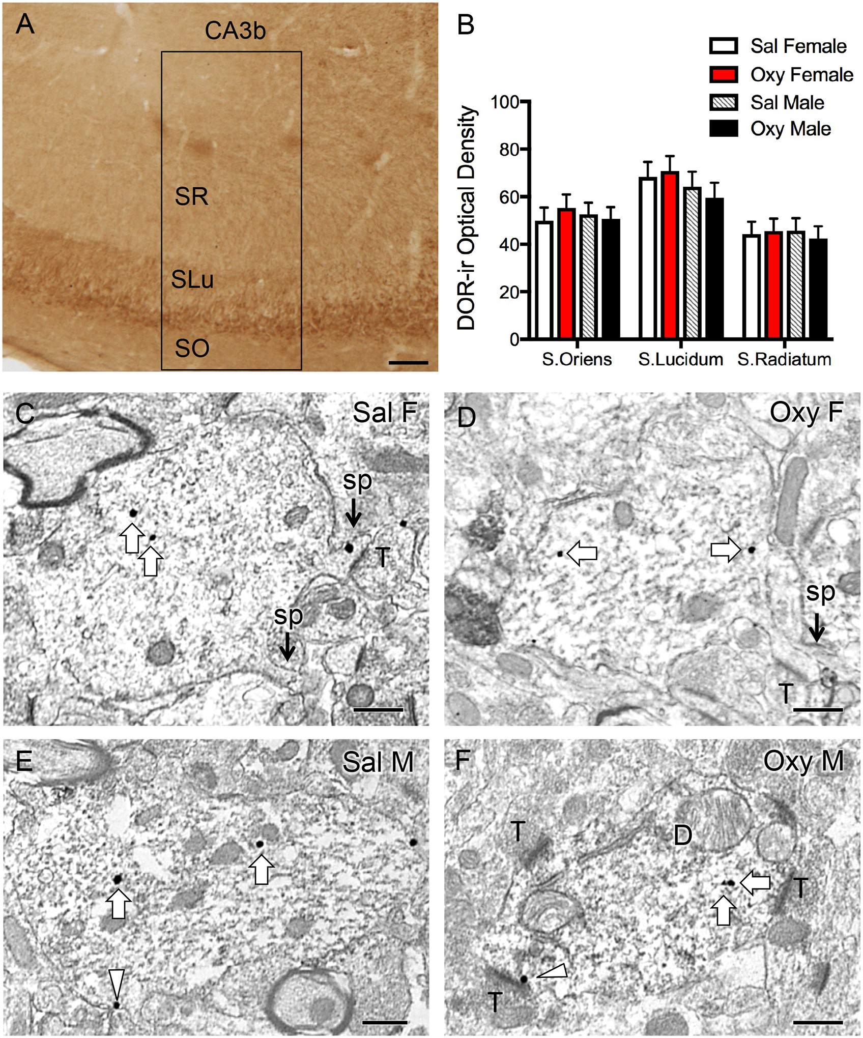
Fig. 4. Representative light and electron micrographs of delta opioid receptor (DOR) in CA3 pyramidal cell dendrites from Sal- and Oxy-CIS female and male rats.

A. Low magnification photomicrograph shows DOR-ir in the CA3 region of the dorsal hippocampus; box indicates area of CA3b sampled for densitometry and EM studies. SO, stratum oriens; SLu, stratum lucidum; SR, stratum radiatum. B. There is no significant difference in DOR-ir levels in any of the subregions of CA3b between Sal-CIS and Oxy-CIS female and male rats. N = 6 rats per group. C-F. Electron micrographs show the distribution of DOR-SIG particles within CA3 pyramidal cell dendrites from a Sal-CIS female (C), an Oxy-CIS female (D), a Sal-CIS male (E), and an Oxy-CIS male (F) rat. Examples of near plasmalemmal (triangle) and cytoplasmic (arrow) DOR-SIG particles in dendrites are shown. CA3 dendrites often have spines (sp) that are contacted by terminals (T; example C). Scale bars: 100 μm (A); 500 nm (C-F).
