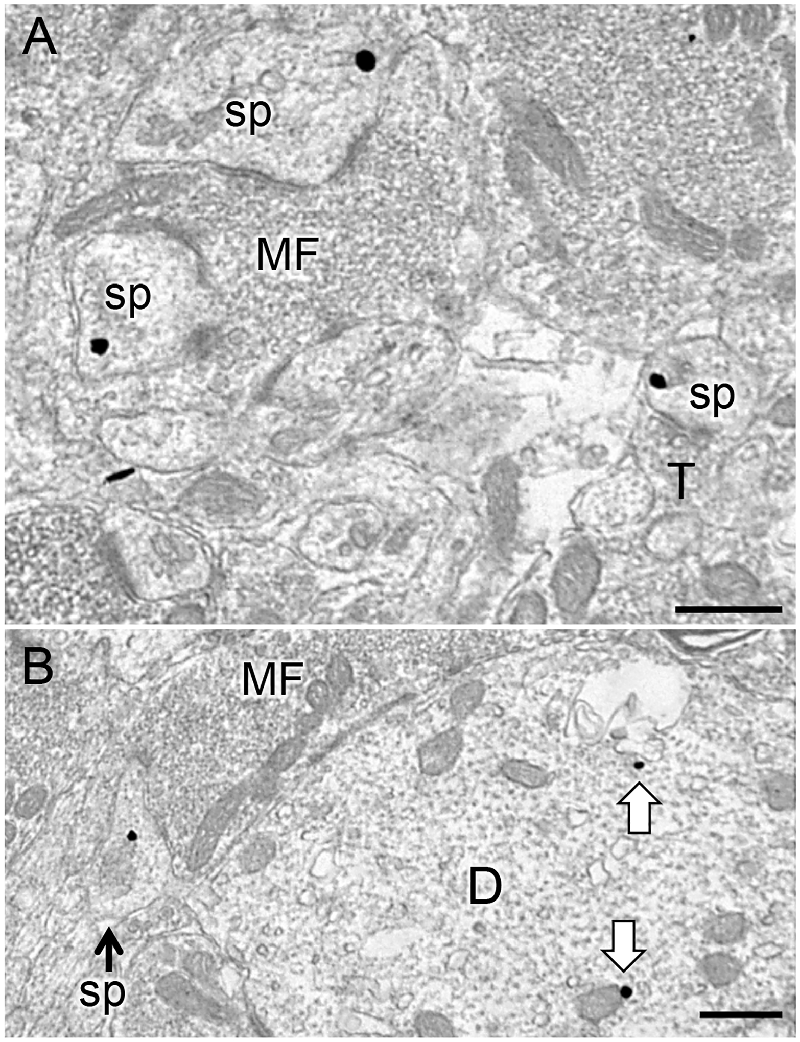
Fig. 6. Representative electron micrographs of DOR-labeled dendritic spines in SLu of CA3.

A. DOR-SIG particles in the cytoplasm of two dendritic spines (sp) in contact with a mossy fiber (MF) in the CA3. Nearby, a DOR-SIG particle is found near the plasmalemma of a spine contacted by a terminal (T; bottom right). B. A DOR-SIG labeled spine emanates from a pyramidal cell dendrite with DOR-SIG particles in the cytoplasm (arrows). A MF abuts the DOR-labeled dendrite (D). Electron micrographs are from Oxy-CIS females. Scale bars: 500 nm.
