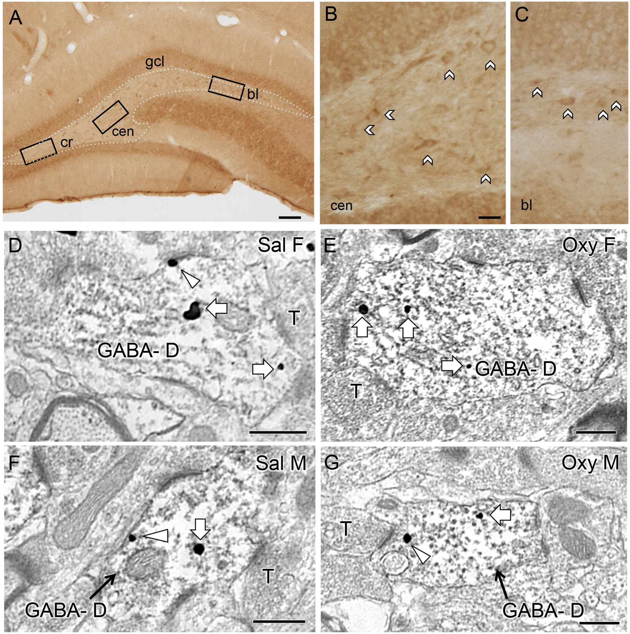
Fig. 7. Representative light and electron micrographs of delta opioid receptor (DOR) in the hilus of the dentate gyrus from Sal- and Oxy-CIS female and male rats.

A. Low magnification photomicrograph shows DOR-containing cells in three regions of the hilus in a CIS female rat (cr = crest; cen = central hilus; bl = blade). B,C. High magnification photomicrographs of the central hilus (B) and dorsal blade (C) show samples of DOR-labeled cells. No differences were observed in the number of DOR-labeled cells in any region of the hilus between Sal-CIS and Oxy-CIS female and male rats (see Table 2). D-G. Electron micrographs show the distribution of DOR-SIG particles within GABA-labeled dendrites (GABA-D) in the hilus from a Sal-CIS female (D), an Oxy-CIS female (E), a Sal-CIS male (F) and an Oxy-CIS male (G) rat. Examples of near plasmalemmal (triangle) and cytoplasmic (arrow) DOR-SIG particles in dendrites are shown. Scale bars: 100 μm (A); 50 μm (B); 500 nm (D-G).
