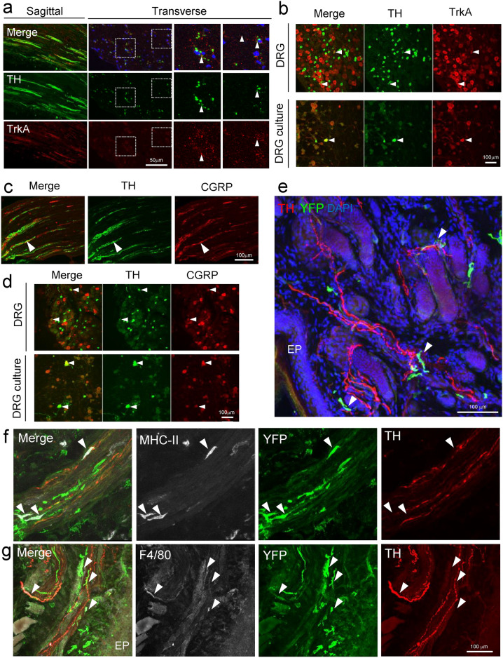
Fig. 4.
SNX25-expressing dermal macrophages are closely associated with TH-positive fibers. (a) Confocal images of the sciatic nerve of WT mice, stained with anti-TH and TrkA antibody. The left panels are a sagittal section and the right panels are a transverse section. Arrowheads denote TH+ TrkA+ nerves. (b) Confocal images of the DRG of WT mice, stained with anti-TH and TrkA antibody. The upper panels are DRGs and the lower panels are a primary DRG neurons. Arrowheads denote TH+ TrkA+ cells. (c) Confocal images of the sciatic nerve of WT mice, stained with anti-CGRP and TH antibody. Arrowheads denote CGRP+ TH+ cells. (d) Confocal images of the DRG of WT mice and primary DRG neurons, stained with anti-CGRP and TH antibody. Arrowheads denote CGRP+ TH+ cells. (e) Confocal images of hind paw skin (naïve) stained for YFP (green) and TH (red) in 4-OHT-treated Cx3cr1CreER; Snx25fl/fl; Ai32 mice. Arrowheads denote nerve-associated YFP+ cells. EP, epidermis. (f) Confocal images of hind paw skin (naïve) stained for YFP (green), MHC-II (white), and TH (red) in 4-OHT-treated Cx3cr1CreER; Snx25fl/fl; Ai32 mice. Arrowheads denote nerve-associated YFP+ MHC-II+ cells. (g) Confocal images of hind paw skin (naïve) stained for YFP (green), F4/80 (white), and TH (red) in 4-OHT-treated Cx3cr1CreER; Snx25fl/fl; Ai32 mice. EP, epidermis. Arrowheads denote nerve-associated YFP+ F4/80+ cells.

