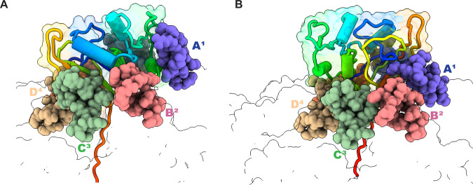
Fig. 2. Fully engaged DHFR can assume distinct orientations with respect to ClpX6 and its RKH loops.
ClpX is shown in outline representation with the positions of subunits A1, B2, C3, and D4 marked and the RKH loops of these subunits shown as spheres in different colors. The DHFR substrate is depicted in cartoon/outline representation in a rainbow-color scheme, with blue representing the N-terminus and red the C-terminus. A DHFR•MTX positioning in the branched-degron structure. B DHFR•MTX positioning in the linear-degron structure.

