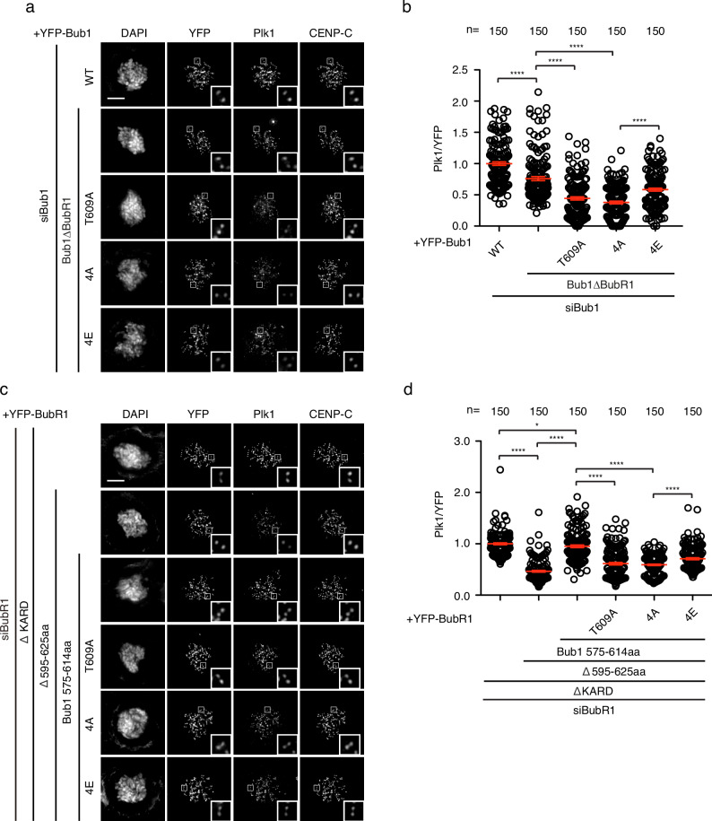
Fig. 4. Bub1-Plk1 interaction requires multiple phosphorylation.
a Representative images of mitotic cells transfected with siRNA oligos against Bub1 and RNAi-resistant constructs expressing YFP-Bub1. The cells were released from RO3306 into the medium with nocodazole (200 ng/ml) for 45 min before fixation and staining with corresponding antibodies. Scale bar is 10 µm. b Quantification of kinetochore signals of Plk1 against YFP-Bub1 from (a). Mean values of 150 kintochores from 10 cells for each condition were presented. The red line indicates the mean value which was set to 1 for wild type Bub1 (WT) sample and the rest was normalized to it. Bar is standard error of the mean. Mann–Whitney U-test was applied. ****P < 0.0001. c Representative images of mitotic cells transfected with siRNA oligos against BubR1 and RNAi-resistant constructs expressing YFP-BubR1. The cells were released from RO3306 into the medium with nocodazole (200 ng/ml) for 45 min before fixation and staining with corresponding antibodies. Scale bar is 10 µm. d Quantification of kinetochore signals of Plk1 against YFP-BubR1 from (c). Mean values of 150 kintochores from 10 cells for each condition were presented. The red line indicates the mean value which was set to 1 for BubR1 ΔKARD sample and the rest was normalized to it. Bar is standard error of the mean. Mann–Whitney U-test was applied. *P < 0.1; ****P < 0.0001.

