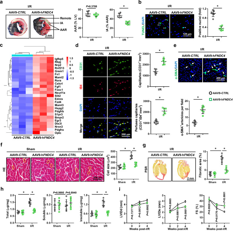
Fig. 2. Cardiac-specific FNDC4 overexpression facilitates cardiomyocyte survival and angiogenesis during cardiac I/R injury.
a Evans blue and TTC double staining was performed to identify the infarct area (IA), area at risk (AAR) and remote area of heart samples 24 h after I/R surgery (n = 6). b Heart samples with or without FNDC4 overexpression were collected 24 h after I/R surgery and subjected to TUNEL staining, and TUNEL+ nuclei were quantified. Arrows indicate TUNEL+ nuclei (n = 6). c Heart samples with or without FNDC4 overexpression were collected 24 h after I/R surgery and subjected to unbiased transcriptome analysis, and the expressions of angiogenesis-related genes were presented using a heatmap (n = 3). d, e Heart samples with or without FNDC4 overexpression were collected 4 weeks after I/R surgery and subjected to immunofluorescence staining, and the numbers of capillaries as well as arterioles were quantified. Arrows indicate α-SMA+ arterioles (n = 6). f, g Heart samples with or without FNDC4 overexpression were collected 4 weeks after I/R surgery and subjected to hematoxylin-eosin (HE) and picrosirius red (PSR) staining, and cell size as well as fibrotic area were quantified. Circles indicate the cross-sectional area of cardiomyocytes (n = 6). h Total, soluble and insoluble collagen content in the heart 4 weeks post-I/R surgery (n = 6). i Cardiac function of FNDC4-overexpressed and control mice was analyzed by transthoracic echocardiography at the indicated time points (n = 6). Data were presented as the mean ± S.D., and analyzed using an unpaired two-tailed Student′s t-test. For the analysis in (f–h), one-way ANOVA followed by Tukey post hoc test was used. For the analysis in (i), repeated measures ANOVA was performed. *P < 0.0001. Source data are provided as a Source Data file.

