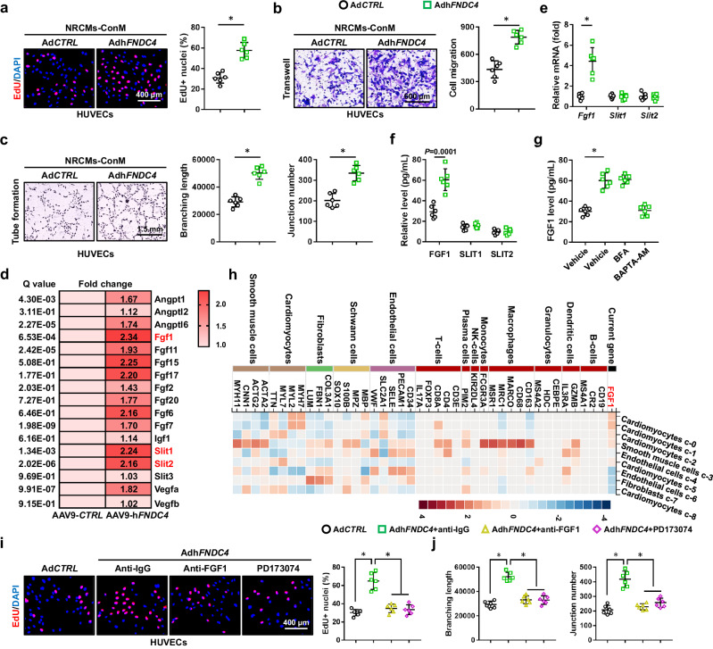
Fig. 5. FNDC4 promotes angiogenesis of endothelial cells through increasing FGF1 secretion from cardiomyocytes.
a Human umbilical vein endothelial cells (HUVECs) were cultured with the conditioned medium (NRCMs-ConM) for 24 h, and then EdU+ nuclei were quantified using a commercial kit (n = 6 independent experiments). b HUVECs were cultured with NRCMs-ConM for 24 h, and then exposed to transwell assay. After 12 h, cells in the lower chamber were stained with crystal violet to quantify the migrated cells (n = 6 independent experiments). c HUVECs were cultured with NRCMs-ConM for 24 h, and then exposed to tube formation assay. After 8 h, the branching length and junction number were quantified (n = 6 independent experiments). d The upregulated angiogenic factors in FNDC4-overexpressed hearts were analyzed using the transcriptome data (n = 3). e The mRNA levels of fibroblast growth factor 1 (Fgf1), slit guidance ligand 1 (Slit1) and Slit2 in NRCMs with or without FNDC4 overexpression (n = 6 independent experiments). f The levels of FGF1, SLIT1 and SLIT2 in the medium of NRCMs with or without FNDC4 overexpression (n = 6 independent experiments). g The level of FGF1 in the medium of NRCMs with brefeldin A (BFA) or BAPTA-AM treatment (n = 6 independent experiments). h Single-cell sequencing data of FGF1 in human hearts. i HUVECs were cultured with NRCMs-ConM in the presence of anti-FGF1 or PD173074 for 24 h, and then EdU+ nuclei were quantified using a commercial kit (n = 6 independent experiments). j HUVECs were cultured with NRCMs-ConM in the presence of anti-FGF1 or PD173074 for 24 h, and then exposed to tube formation assay (n = 6 independent experiments). Data were presented as the mean ± S.D., and analyzed using an unpaired two-tailed Student′s t-test. For the analysis in (g–j), one-way ANOVA followed by Tukey post hoc test was used. *P < 0.0001. Source data are provided as a Source Data file.

