
Fig. 1. Confocal Image Analysis Methodology.
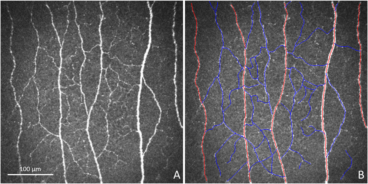
Illustration of confocal image analysis methodology can be observed in the in vivo confocal image provided to each grader (A) and in the same image with the nerves traced (B). Red tracings highlight main nerves, and blue tracings highlight branch nerves.