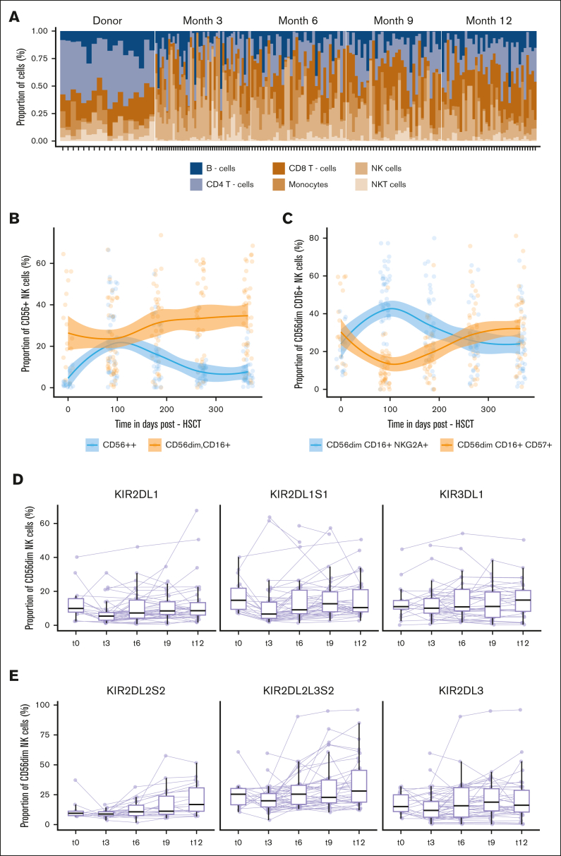
Figure 3.
Immune-cell composition during the first year after HSCT. (A) Relative proportions of 6 major cell populations: CD8+ and CD4+ T cells, NK T cell, NK cell, monocytes, and B cells assessed at the indicated time points after HSCT: t0 (n = 18), t3 (n = 44), t6 (n = 35), t9 (n = 32), and t12 (n = 37). Each stacked bar represents an individual. (B) Evolution of the frequency of CD56bright, CD56dim, CD16+ NK cells at indicated time points after HSCT: t0 (n = 18), t3 (n = 44), t6 (n = 35), t9 (n = 32), and t12 (n = 37) fitted using LOESS regression with a 95% confidence interval. Day 0 indicates a pre-HSCT state of the donor. Color-coded lines represent linear regression according to cell subset. (C) Evolution of the frequency of CD56dim, CD16+, NKG2A+, CD57neg and CD56dim, CD16+, NKG2Aneg, and CD57+ NK cells at the indicated time points after HSCT: t0 (n = 18), t3 (n = 44), t6 (n = 35), t9 (n = 32), and t12 (n = 37) fitted using LOESS regression with a 95% confidence interval. Day 0 indicates a pre-HSCT state of the donor. Color-coded lines represent linear regression according to the cell subset. (D, E) Proportion of KIR+ CD56dim NK cell at the indicated time points after HSCT: t0 (n = 18), t3 (n = 44), t6 (n = 35), t9 (n = 32), and t12 (n = 37). The lines connect paired samples. Box plots display medians and IQRs, with whiskers representing 1.5× IQR.

