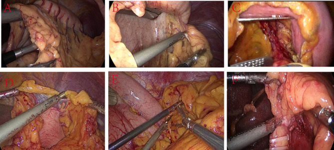
Fig. 3.
Comparison of view and instrument placement between SSP SILS + 1 and CLS. A-C: SSP SILS + 1; D-F: CLS. (A) Upon exposing the greater omentum, the FLFSF was placed posteriorly to the stomach to lift the stomach through the omental opening, while also pulling the omentum to expose the surgical field; this maneuver facilitated the use of the ultrasonic scalpel to detach the omentum; (B) FLFSF were used to lift the stomach in the upper right direction, thereby exposing the short gastric vessels; (C) After ligating the left gastric artery, FLFSF was used to lift the stomach, stretch and expose the left gastric artery, which was excised with a linear cutting stapler; (D) The surgeon and assistant collaborated to disconnect the omentum with the assistance of the first assistant; this procedure involved the coordinated effort of three surgons; (E) Routine five hole exposure of severed gastric short blood vessels requiring an assistant to use FLFSF to individually lift the liver to expose the surgical field; (F) The CLS procedure exposing the severed left gastric blood vessel, while an assistant using FLFSF to individually lift the liver to expose the surgical field.

