Abstract
Background:
One factor contributing to rotational deformity of the first metatarsal in hallux valgus is torsion of the metatarsal itself. Hallux rigidus also involves reduction of the longitudinal arch, but metatarsal torsion has not been discussed. We hypothesized that metatarsal torsion may be a morphologic change unique to hallux valgus. We compared 3-dimensional (3D) torsion of the first to fifth metatarsals between feet with hallux valgus, feet with hallux rigidus, and healthy control feet to investigate differences in the effects on pathologic conditions.
Methods:
Participants were women of East Asian descent. There were 16, 16, and 14 feet in the control, hallux valgus, and hallux rigidus groups, respectively. One randomly selected control foot was designated as the reference foot. For comparison, nonweightbearing computed tomography images of the metatarsals were reconstructed in 3D, and the proximal and distal areas were superimposed on the reference foot. Torsion angle was defined as the rotational angle of the distal part of the articular axis relative to the proximal area. In the hallux valgus group, correlations of torsion angle with hallux valgus angle and intermetatarsal angle were calculated.
Results:
The hallux valgus group had greater average pronation torsion in the first metatarsal than the control group and hallux rigidus group (11 and 13 degrees greater, respectively, P < .01). No significant differences were observed for the second to fifth metatarsals (P > .05). There was no significant correlation with hallux valgus angle or first-second intermetatarsal angle in the hallux valgus group (P > .05).
Conclusion:
Hallux valgus feet had pronation deformities in the first metatarsals not observed in control or hallux rigidus feet, meaning that torsion toward pronation (eversion) in the first metatarsal was unique to hallux valgus. Improved surgical correction to diminish pronation may be necessary in patients with hallux valgus patients because of first metatarsal pronation in the first tarsometatarsal to normalize mechanical first-ray alignment.
Level of Evidence: Level III, case-control stud.
Keywords: metatarsal, torsion, hallux valgus, hallux rigidus, computed tomography, 3D
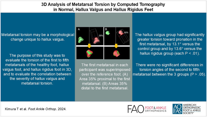
Visual Abstract.
This is a visual representation of the abstract.
Introduction
Hallux valgus is a common foot disorder that affects 30% of people aged ≥65 years. 16 Hallux valgus is a 3-dimensional (3D) deformity, with deformities in the sagittal, transverse, and frontal planes, not simply valgus of the hallux.2,6,12 -15 In general, the first metatarsal head is considered to be in a pronated position in the hallux valgus foot, and computed tomography (CT) studies have been conducted.2,3 Recently, it has been suggested that correction of the rotation of the first metatarsal head is necessary to achieve a good corrective position in hallux valgus treatment, and it is also closely related to correction of the sesamoid position.17,22
Rotation of the plantar surface in hallux valgus at the first metatarsophalangeal joint is caused by rotation of the first metatarsal at the first tarsometatarsal joint and mobility at the tarsometatarsal joint, which lowers the arch and causes the entire foot to collapse inward. In addition, torsion of the first metatarsal is also a factor. 18 Because 2D evaluation of this torsion on plain radiographs is difficult, few studies have evaluated torsion of the first metatarsal.5,18 Also, there have been no reports on torsion of the second to fifth metatarsals in hallux valgus or hallux rigidus feet.
Hallux rigidus, on the other hand, is a degenerative condition of the metatarsophalangeal joint of the hallux with pain and is characterized by progressive osteophyte formation and restricted range of motion. 10 Deformity of the first ray is a feature common to hallux rigidus and hallux valgus.
Although the commonalities of the pathophysiology have been reported, 11 such as hypermobility of the first ray, the final deformity is different.
We hypothesized that there is torsion of the first metatarsal toward pronation in the hallux valgus foot, but not in the hallux rigidus foot. The purpose of this study was to evaluate the torsion of the first to fifth metatarsals of the healthy foot, hallux valgus foot, and hallux rigidus foot in 3D, and to evaluate the correlation between the severity of hallux valgus and metatarsal torsion.
Materials and Methods
Participants
In this case-control study, we examined 28 feet of 15 healthy volunteers with no history or symptoms of foot disorders (control group), 30 feet of 24 patients with hallux valgus (HV group), and 20 feet of 11 patients with hallux rigidus (HR group). All participants were women of East Asian descent, as classified in electronic health records. The HV group consisted of patients who visited our clinic between June 2016 and June 2023 at Jikei university hospital, Tokyo, Japan. We excluded control and patients with hallux valgus who exhibited some degenerative changes. The HR group consisted of patients who visited our clinic for hallux pain between June 2018 and June 2023 and were diagnosed as having HR based on plain radiographs and clinical symptom by an orthopaedic surgeon with about 30 years of specialization in foot and ankle surgery (M.K.). The participants in the control group were recruited from among the staff and students at our university. In both the HV and HR groups, we excluded patients with inflammatory arthritis such as rheumatism or another foot condition.
The Research Ethics Committee of our institution approved this study, which complied with the Declaration of Helsinki. All participants provided written informed consent.
Imaging and Image Analysis
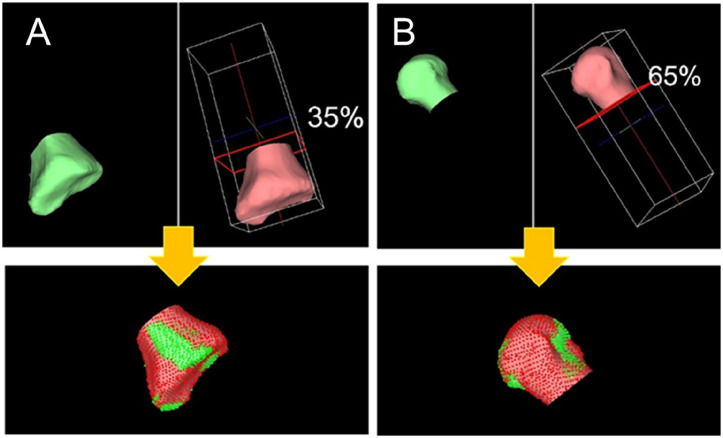
Weightbearing plain radiographs and foot and ankle CT imaging (0.75-mm slices) was performed for all 3 groups (HV, HR, and control). The hallux valgus angle, the first-second intermetatarsal angle (IMA 1-2) were measured on weightbearing plain radiographs of all subjects. In the HR group, evaluation of weightbearing plain radiographs was performed according to the Hattrup and Johnson radiographic classification of hallux rigidus. 8 One randomly selected foot from the control group was designated as the reference foot. For comparisons between groups, each metatarsal was semiautomatically segmented, and a 3D model was generated using image analysis software (Mimics ver. 21; Materialise, Leuven, Belgium), and 35% of the proximal and distal areas were taken and superimposed on the reference foot using the iterative closest point algorithm9,20,21 (Figure 1), which allows superimposition of 3D objects without specifying anatomical landmarks. The torsion angle was defined as the angle of rotation of the distal part of the articular axis relative to the proximal area compared with the reference foot. In the hallux valgus group, correlations of the torsion angle with the hallux valgus angle and intermetatarsal angle were also calculated.
Figure 1.
The first metatarsal in each participant was superimposed over the reference foot. (A) Area 35% proximal to the first metatarsal. (B) Areas 35% distal to the first metatarsal.
Statistical Analysis
A power analysis was performed to determine the minimum number of patients needed for each group. The sample size was estimated for unpaired 2-tailed t test. Ota et al 18 reported the first metatarsal torsional angle as 17.6 ± 7.7 degrees toward pronation in a hallux valgus group and 4.7 ± 4.0 degrees toward pronation for a healthy control group. For these 2 averages (effect size = 1.500, setting alpha = 0.0167, and 1 − beta = 0.80), the power was calculated using a sample-size calculation tool (G*Power, version 3.0.10; Franz Faul, University of Kiel, Kiel, Germany), and a minimum of 11 feet were required for each group, for a total of 33 feet. We therefore set a sample size of more than 11 feet for each group. Continuous data with a normal distribution are shown as the mean ± standard deviation, and data with a non-normal distribution are shown as median (interquartile range). Normally distributed continuous volume data were compared using 1-way analysis of variance and the unpaired t test, whereas non-normally distributed continuous volume data were compared using the Kruskal-Wallis test and the Mann-Whitney U test. To correct for multiplicity of tests, P values were corrected using the Bonferroni method. Spearman rank correlation coefficient was calculated to examine the correlation between the hallux valgus angle, IMA 1-2, and torsion angle of the first to fifth metatarsal (pronation, negative; supination, positive). P <.05 was considered statistically significant. Analysis was performed using SPSS version 22.0 for Windows (IBM Japan, Tokyo, Japan).
Results
All patients were female. Age and body mass index for each group are shown in Table 1. There were no significant differences in these parameters between the 3 groups (all P > .05).
Table 1.
Characteristics of Participants by Group.
| (1) Control | (2) Hallux Valgus | (3) Hallux Rigidus | P Value | |||||||
|---|---|---|---|---|---|---|---|---|---|---|
| n | Data | n | Data | n | Data | P Value (for All) | (1) vs (2) | (1) vs (3) | (2) vs (3) | |
| Number of feet | 28 | 30 | 20 | |||||||
| Female, n | 15 | 24 | 11 | |||||||
| Age, y, mean ± SD | 15 | 54.4 ± 6.4 | 24 | 59.6 ± 14.3 | 11 | 60.9 ± 11.3 | .131 a | >.999 b | >.999 b | >.999 b |
| BMI, mean ± SD | 15 | 20.6 ± 2.5 | 24 | 21.6 ± 3.5 | 11 | 22.7 ± 2.5 | .229 a | >.999 b | >.999 b | >.999 b |
Abbreviation: BMI, body mass index.
One-way analysis of variance.
Unpaired t test (Bonferroni corrected).
Table 2 shows the results for between-group comparisons of hallux valgus angle, IMA 1-2, and torsion angle of the first to fifth metatarsal. The torsion angle of the first metatarsal was 1.71 ± 6.76 degrees toward supination in the control group, 11.39 ± 7.35 degrees toward pronation in the hallux valgus group, and 2.18 ± 12.06 degrees toward supination in the hallux rigidus group. The hallux valgus group had significantly greater torsion toward pronation in the first metatarsal, by 13.1 degrees vs the control group and by 13.6 degrees vs the hallux rigidus group (each P < .01). There were no significant differences in torsion angles of the second to fifth metatarsal between the 3 groups (P > .05).
Table 2.
Comparisons Between Groups. a
| (1) Control | (2) Hallux Valgus | (3) Hallux Rigidus | P Value (for All) | P Value (Bonferroni Corrected) | ||||||
|---|---|---|---|---|---|---|---|---|---|---|
| n | Data | n | Data | n | Data | (1) vs (2) | (1) vs (3) | (2) vs (3) | ||
| Hattrup and Johnson radiographic classification | 0 | 0 | 20 | — | — | — | ||||
| I | — | — | 9 | |||||||
| II | — | — | 10 | |||||||
| II | — | — | 1 | |||||||
| Hallux valgus angle, median [IQR] | 28 | 12.0 [12.0, 14.0] | 29 | 38.0 [28.5, 50.0] | 20 | 13.0 [12.0, 14.8] | <.01 b | <.01 c | >.999 c | <.01 c |
| First-second intermetatarsal angle, median [IQR] | 28 | 8.0 [8.0, 9.0] | 29 | 18.0 [13.5, 22.0] | 20 | 8.0 [7.3, 9.0] | <.01 b | <.01 c | >.999 c | <.01 c |
| First metatarsal torsion angle, degrees, mean ± SD | 28 | 1.71 ± 6.76 | 30 | −11.39 ± 7.35 | 20 | 2.18 ± 12.06 | <.01 b | <.01 d | >.999 d | <.01 d |
| Second metatarsal torsion angle, degrees, mean ± SD | 28 | 0.13 ± 8.54 | 30 | −0.26 ± 17.17 | 20 | 2.43 ± 8.76 | .744 e | >.999 d | >.999 d | >.999 d |
| Third metatarsal torsion angle, degrees, mean ± SD | 28 | 0.27 ± 5.57 | 30 | 0.78 ± 8.40 | 20 | −1.26 ± 10.32 | .675 e | >.999 d | >.999 d | >.999 d |
| Fourth metatarsal torsion angle, degrees, mean ± SD | 28 | −0.25 ± 6.36 | 30 | 1.25 ± 12.09 | 20 | −2.79 ± 11.55 | .397 e | >.999 d | >.999 d | .733 d |
| Fifth metatarsal torsion angle, degrees, mean ± SD | 28 | 5.16 ± 11.71 | 30 | −2.01 ± 16.31 | 20 | 2.00 ± 9.83 | .127 e | .183 d | .988 d | .990 d |
Pronation, positive; supination, negative. Data in bold are statistically significant at P < .05.
One-way analysis of variance.
Unpaired t-test (Bonferroni corrected).
Mann-Whitney U test (Bonferroni corrected).
Kruskal-Wallis test.
Table 3 shows correlations between hallux valgus angle, IMA 1-2, and torsion angle of the first to fifth metatarsal in the hallux valgus group. There were no correlations between these parameters (P > .05).
Table 3.
Correlations of Hallux Valgus Angle and First-Second Intermetatarsal Angle With First to Fifth Metatarsal Torsion Angle. a
| Hallux Valgus Angle | First-Second Intermetatarsal Angle | |||
|---|---|---|---|---|
| ρ b | P Value | ρ b | P Value | |
| First metatarsal torsion angle | −0.151 | .249 | −0.249 | .231 |
| Second metatarsal torsion angle | −0.080 | .486 | 0.046 | .692 |
| Third metatarsal torsion angle | 0.017 | .883 | 0.121 | .293 |
| Fourth metatarsal torsion angle | 0.051 | .661 | −0.009 | .938 |
| Fifth metatarsal torsion angle | −0.112 | .332 | −0.185 | .106 |
Pronation, positive; supination, negative.
ρ, Spearman rank correlation coefficient.
Discussion
The findings of this study showed that the first metatarsal has torsion deformity in the pronation direction in the hallux valgus foot compared with normal feet, but not in the hallux rigidus foot, thus supporting our hypothesis.
Although some studies have investigated the first metatarsal pronation angle in hallux valgus,2,4,12 -15,19 only a few reports have discussed metatarsal torsion itself.1,23,24 Cruz et al 5 performed 2D measurements of first metatarsal torsion on CT slices. They reported a torsion angle of 15.4 (range, 1.6-32.5) degrees toward pronation in the hallux valgus group and 3.5 (range, −7.4 to 15.6) degrees toward pronation in the control group. Ota et al 18 examined first metatarsal torsion in 27 patients with hallux valgus and 12 control patients in 3D. The torsional angle of the first metatarsal was 17.6 ± 7.7 degrees toward pronation in the hallux valgus group and 4.7 ± 4.0 degrees toward pronation for the control group. From that study, torsion of the first metatarsal was around 10 degrees toward pronation compared with the control group. These results were similar to ours. However, they did not evaluate the second to fifth metatarsal. In our results, there were no significant differences between the hallux valgus and control groups in the torsion angle of the second to fifth metatarsal. Furthermore, to our knowledge, no studies have evaluated metatarsal torsion in hallux rigidus. No significant differences in metatarsal torsion were found between the control and hallux rigidus groups. However, the present results show that hallux valgus feet had more torsion toward pronation compared with hallux rigidus feet. Our results indicate that first metatarsal torsion toward pronation was a deformity unique to hallux valgus.
Our results also show that the degree of rotation was not correlated with severity. The sesamoid complex and the plantar fascia might create a tendency toward relatively lateral displacement, which would increase the rotation of the first metatarsal and valgus of metatarsophalangeal joint.
In an interesting study on metatarsal torsion in humans, early hominids, and primates, Drapeau and Harmon 7 evaluated metatarsal torsion in monkeys, apes, humans, and australopiths. They reported that the ape foot is characterized by an everted first metatarsal. However, the human foot is characterized by a relatively untwisted first metatarsal. Their results suggest that during human evolution, the first metatarsal twisted toward eversion as the opposing position of the hallux was lost. 7 Given that our results showed that the first metatarsal in hallux valgus feet had torsion deformity toward pronation, phylogenetically, hallux valgus may be considered to be the result of degeneration.
Residual pronation deformity postoperatively may lead to recurrence of hallux valgus. 17 Our results indicate that hallux valgus feet have torsion deformity toward pronation.
This study had some limitations. First, the sample size was small and all participants were Asian women. Therefore, our results may not apply to both sexes or to all ethnicities, statures, or other patient factors. Non-Asians and men should also be analyzed to better understand the pathology of these conditions, and they should also be included in the control group. Also, although the sample size was small, it was determined by an a priori sample size calculation. Second, most patients in the hallux valgus feet had relatively severe deformity, whereas those in the hallux rigidus group had mild to moderate deformity, and this difference may have influenced our results. Third, the participant age distribution was skewed toward 50 years or older. Future studies should include younger patients with hallux valgus and hallux rigidus. However, this may be challenging given that early onset is rare.
Conclusion
In conclusion, by using CT images and a 3D analysis system with the iterative closest point algorithm, we were able to evaluate metatarsal torsion in detail for hallux valgus, hallux rigidus, and normal feet. The hallux valgus feet had significantly greater torsion toward pronation in the first metatarsal compared with the hallux rigidus and normal feet. However, no correlation was found between severity of hallux valgus and torsion angle. Improved surgical correction to diminish pronation may be necessary in patients with hallux valgus patients because of first metatarsal pronation in the first tarsometatarsal to normalize mechanical first-ray alignment.
Supplemental Material
Supplemental material, sj-pdf-1-fao-10.1177_24730114241294074 for 3D Analysis of Metatarsal Torsion by Computed Tomography in Normal, Hallux Valgus, and Hallux Rigidus Feet by Tadashi Kimura, Makoto Kubota, Takumi Kihara, Naoki Suzuki, Asaki Hattori and Mitsuru Saito in Foot & Ankle Orthopaedics
Footnotes
Ethical Approval: Ethical approval for this study was obtained from the Ethics Committee of the Jikei University School of Medicine for Biomedical Research (approval no. 25-113(7248)).
The author(s) declared no potential conflicts of interest with respect to the research, authorship, and/or publication of this article. Disclosure forms for all authors are available online.
Funding: The author(s) disclosed receipt of the following financial support for the research, authorship, and/or publication of this article: This work was supported by the Grants-in-Aid for Scientific Research of Japan Society for the Promotion of Science, Grant Number JP22K16776.
ORCID iD: Tadashi Kimura, MD, PhD,  https://orcid.org/0000-0002-9812-0697
References
- 1. Bakshi N, Steadman J, Philippi M, et al. Association between hindfoot alignment and first metatarsal rotation. Foot Ankle Int. 2021;43(1):105-112. doi: 10.1177/10711007211033514 [DOI] [PubMed] [Google Scholar]
- 2. Campbell B, Miller MC, Williams L, Conti SF. Pilot study of a 3-dimensional method for analysis of pronation of the first metatarsal of hallux valgus patients. Foot Ankle Int. 2018;39(12):1449-1456. doi: 10.1177/1071100718793391 [DOI] [PubMed] [Google Scholar]
- 3. Collan L, Kankare JA, Mattila K. The biomechanics of the first metatarsal bone in hallux valgus: a preliminary study utilizing a weight bearing extremity CT. Foot Ankle Surg. 2013;19(3):155-161. doi: 10.1016/j.fas.2013.01.003 [DOI] [PubMed] [Google Scholar]
- 4. Conti MS, Patel TJ, Caolo KC, et al. Correlation of different methods of measuring pronation of the first metatarsal on weightbearing CT scans. Foot Ankle Int. 2021;42(8):1049-1059. doi: 10.1177/10711007211003090 [DOI] [PubMed] [Google Scholar]
- 5. Cruz EP, Wagner FV, Henning C, Sanhudo JAV, Pagnussato F, Galia CR. Does hallux valgus exhibit a deformity inherent to the first metatarsal bone? J Foot Ankle Surg. 2019;58(6):1210-1214. doi: 10.1053/j.jfas.2018.09.031 [DOI] [PubMed] [Google Scholar]
- 6. Dayton P, Kauwe M, DiDomenico L, Feilmeier M, Reimer R. Quantitative analysis of the degree of frontal rotation required to anatomically align the first metatarsal phalangeal joint during modified tarsal-metatarsal arthrodesis without capsular balancing. J Foot Ankle Surg. 2016;55(2):220-225. doi: 10.1053/j.jfas.2015.08.018 [DOI] [PubMed] [Google Scholar]
- 7. Drapeau MS, Harmon EH. Metatarsal torsion in monkeys, apes, humans and australopiths. J Hum Evol. 2013;64(1):93-108. doi: 10.1016/j.jhevol.2012.10.008 [DOI] [PubMed] [Google Scholar]
- 8. Hattrup SJ, Johnson KA. Subjective results of hallux rigidus following treatment with cheilectomy. Clin Orthop Relat Res. 1988;226:182-191. [PubMed] [Google Scholar]
- 9. Kimura T, Kubota M, Taguchi T, Suzuki N, Hattori A, Marumo K. Evaluation of first-ray mobility in patients with hallux valgus using weight-bearing CT and a 3-D analysis system: a comparison with normal feet. J Bone Joint Surg Am. 2017;99(3):247-255. doi: 10.2106/JBJS.16.00542 [DOI] [PubMed] [Google Scholar]
- 10. Lucas DE, Hunt KJ. Hallux rigidus: relevant anatomy and pathophysiology. Foot Ankle Clin. 2015;20(3):381-389. doi: 10.1016/j.fcl.2015.04.001 [DOI] [PubMed] [Google Scholar]
- 11. Maceira E, Monteagudo M. Functional hallux rigidus and the Achilles-calcaneus-plantar system. Foot Ankle Clin. 2014;19(4):669-699. doi: 10.1016/j.fcl.2014.08.006 [DOI] [PubMed] [Google Scholar]
- 12. Najefi AA, Alsafi MK, Malhotra K, Patel S, Cullen N, Welck M. Repeatability of weightbearing computed tomography measurement of first metatarsal alignment and rotation. Foot Ankle Int. 2021;43(2):260-266. doi: 10.1177/10711007211035387 [DOI] [PubMed] [Google Scholar]
- 13. Najefi AA, Katmeh R, Zaveri AK, et al. Imaging findings and first metatarsal rotation in hallux valgus. Foot Ankle Int. 2022;43(5):665-675. doi: 10.1177/10711007211064609 [DOI] [PubMed] [Google Scholar]
- 14. Najefi AA, Malhotra K, Patel S, Cullen N, Welck M. Assessing the rotation of the first metatarsal on computed tomography scans: a systematic literature review. Foot Ankle Int. 2021;43(1):66-76. doi: 10.1177/10711007211020676 [DOI] [PubMed] [Google Scholar]
- 15. Najefi AA, Zaveri A, Alsafi MK, et al. The assessment of first metatarsal rotation in the normal adult population using weightbearing computed tomography. Foot Ankle Int. 2021;42(10):1223-1230. doi: 10.1177/10711007211015187 [DOI] [PubMed] [Google Scholar]
- 16. Nix S, Smith M, Vicenzino B. Prevalence of hallux valgus in the general population: a systematic review and meta-analysis. J Foot Ankle Res. 2010;3:21. doi: 10.1186/1757-1146-3-21 [DOI] [PMC free article] [PubMed] [Google Scholar]
- 17. Okuda R, Kinoshita M, Yasuda T, Jotoku T, Kitano N, Shima H. Postoperative incomplete reduction of the sesamoids as a risk factor for recurrence of hallux valgus. J Bone Joint Surg Am. 2009;91(7):1637-1645. doi: 10.2106/JBJS.H.00796 [DOI] [PubMed] [Google Scholar]
- 18. Ota T, Nagura T, Kokubo T, et al. Etiological factors in hallux valgus, a three-dimensional analysis of the first metatarsal. J Foot Ankle Res. 2017;10:43. doi: 10.1186/s13047-017-0226-1 [DOI] [PMC free article] [PubMed] [Google Scholar]
- 19. Randich JR, John KJ, Gomez K, Bush WJ. Frontal plane rotation of the first ray in hallux valgus using standing CT. J Foot Ankle Surg. 2021;60(3):489-493. doi: 10.1053/j.jfas.2020.05.022 [DOI] [PubMed] [Google Scholar]
- 20. Robb RA. The biomedical imaging resource at Mayo Clinic. IEEE Trans Med Imaging. 2001;20(9):854-867. doi: 10.1109/42.952724 [DOI] [PubMed] [Google Scholar]
- 21. Robb RA, Hanson DP, Karwoski RA, Larson AG, Workman EL, Stacy MC. Analyze: a comprehensive, operator-interactive software package for multidimensional medical image display and analysis. Comput Med Imaging Graph. 1989;13(6):433-454. doi: 10.1016/0895-6111(89)90285-1 [DOI] [PubMed] [Google Scholar]
- 22. Shibuya N, Kyprios EM, Panchani PN, Martin LR, Thorud JC, Jupiter DC. Factors associated with early loss of hallux valgus correction. J Foot Ankle Surg. 2018;57(2):236-240. doi: 10.1053/j.jfas.2017.08.018 [DOI] [PubMed] [Google Scholar]
- 23. Steadman J, Bakshi N, Arena C, Leake R, Barg A, Saltzman CL. Normative distribution of first metatarsal axial rotation. Foot Ankle Int. 2021;42(8):1040-1048. doi: 10.1177/10711007211001015 [DOI] [PubMed] [Google Scholar]
- 24. Steadman J, Barg A, Saltzman CL. First metatarsal rotation in hallux valgus deformity. Foot Ankle Int. 2021;42(4):1071100721997149. doi: 10.1177/1071100721997149 [DOI] [PubMed] [Google Scholar]
Associated Data
This section collects any data citations, data availability statements, or supplementary materials included in this article.
Supplementary Materials
Supplemental material, sj-pdf-1-fao-10.1177_24730114241294074 for 3D Analysis of Metatarsal Torsion by Computed Tomography in Normal, Hallux Valgus, and Hallux Rigidus Feet by Tadashi Kimura, Makoto Kubota, Takumi Kihara, Naoki Suzuki, Asaki Hattori and Mitsuru Saito in Foot & Ankle Orthopaedics


