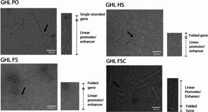
Fig. 3.
Verification of the structural integrity of ultracentrifugation-filtered DNA nanoparticles by transmission electron microscopy (TEM). A representative DNA nanoparticle with key structural elements is located to the lower right of each representative TEM field. The black arrows point to the representative DNA nanoparticles within the TEM field. ssDNA and loosely crosslinked domains appear as relatively amorphous consolidated networks whereas linear duplexes containing the promoter and enhancer appear as curved tails due to the enhanced persistence length of dsDNA relative to ssDNA31. Compact, highly crosslinked origami regions display the canonical multi-helix particle morphology. Scale bar: 50 nm.

