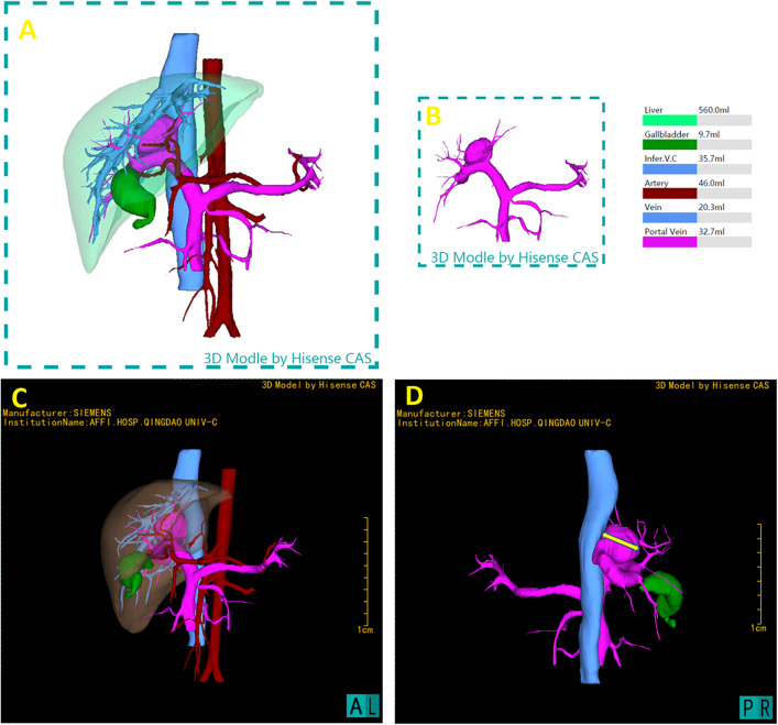
Figure 2.
3D reconstruction utilizing the hisense computer-assisted surgery (CAS) system and preoperative three-dimensional model. (A) Full reconstruction of liver and surrounding vasculature, showing liver in transparent, measured 560.0 ml (B) portal vein measured 32.7 ml, including the aneurysm where the shunt located. (C) Represent the lateral perspectives of the 3D reconstruction, showcasing the liver in a semi-transparent state to clarify its internal anatomical structures and vascular variances. (D) The measurement of the maximum transverse diameter of the portal vein's tumor-like dilatation, measured as 26.36 mm, providing critical spatial dimensions.

