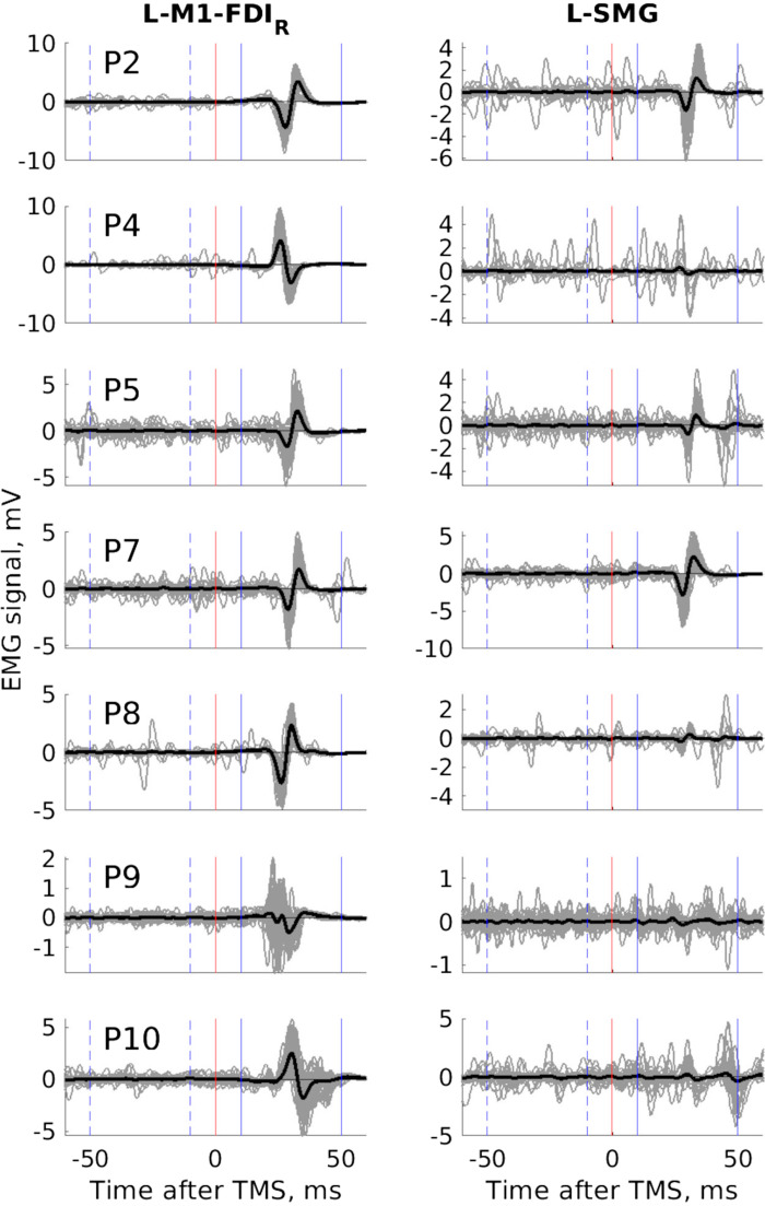
Figure 3.
Transcranial magnetic stimulation (TMS, biphasic) over the left supramarginal gyrus (L-SMG) produces motor-evoked potentials (MEPs) in the right first dorsal interosseous (FDIR) muscle during the pegboard task (experiment 2b, N = 7). Each panel shows all 60 raw traces (thin gray lines) and the grand average mean electromyographic (EMG) signal (thick black lines) after TMS over the primary motor cortex (L-M1-FDIR, left) and L-SMG (right) from each participant (from top to bottom, P2, 4, 5, 7, 8, 9, 10). With TMS over SMG, all 7 participants show TMS-related EMG deflections in the 10 ms to 50 ms window following TMS, some participants more than others, all participants more in the post-TMS than the pre-TMS window. The y-axis scale varies from a minimum of ±1 mV to ±10 mV.

