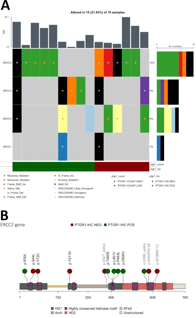
Fig. 3.

A: Oncoplot showing ERCC1-4, non-mutated cases are filtered out, of the 70 patients, sorted by PTGR1 positive and PTGR1 negative (by both IHC and RNA expression). Variants known pathogenicity by OncoKB is marked with a colored dot. B: mapping the mutations of ERCC2 along the gene, depicting the helicases and genomic structures. Missense mutations in the helicase domain are considered pathogenic, all missense mutations are within the helicase domains. Splice site and frameshift mutations are noted in light-grey text. Each dot is colored by PTGR1 IHC status.
