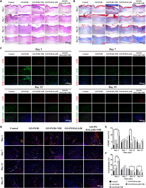
Fig. 7.

Histological evaluation of wound healing quality. (A) H&E staining and (B) Masson staining images of skin on 3, 7, 12, and 19 d. Scale bar, 500 μm. (C) Immunofluorescence staining images of F4/80 and CD206 of skin on 3, 7, 12, and 19 d. Scale bar, 100 μm. (D) Immunofluorescence staining images of CD31 (red) and α-SMA (green) of skin on 3, 7, 12, and 19 d. Scale bar, 100 μm. (E) Statistical value of CD206+ area (n = 4, *P < 0.05, **P < 0.01, ***P < 0.001). (F) Statistical value of vessel density calculated from immunofluorescence staining images (n = 4, *P < 0.05, **P < 0.01, ***P < 0.001).
