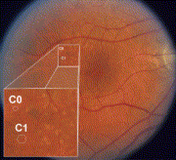
Figure 1.
In an eye with multiple drusen variants, the Age-Related Eye Disease Study drusen grading circles C0 (63-μm diameter) and C1 (125-μm diameter) are superimposed for size comparison. Small drusen are smaller than the C0 circle (drupelets). Lesions larger than C0 but less than C1 are considered medium drusen, and lesions larger than C1 are large drusen. Within the inset, drupelets and medium drusen are seen. Faint reticular drusen also may be seen in the superior macular region.

