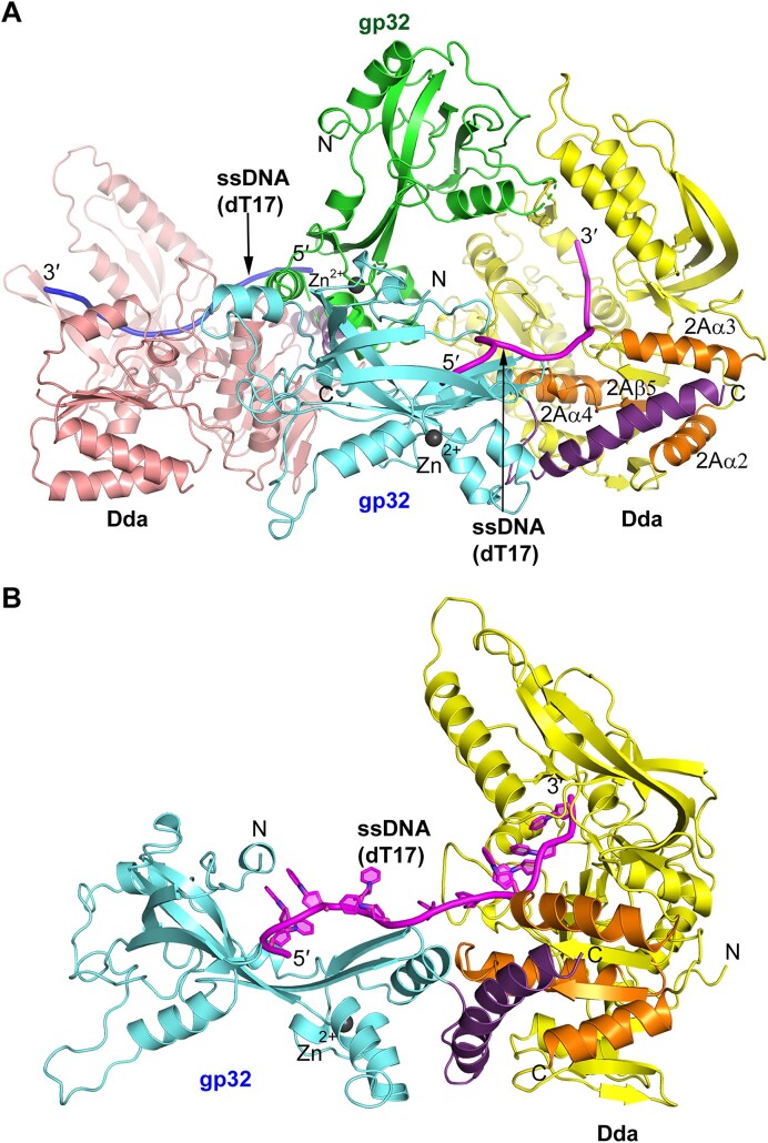
Figure 1.
Crystal structure of the gp32–Dda–dT17 complex. (A) The two complexes in the crystal asymmetric unit. The gp32 molecules are shown in green and cyan, and the two Dda molecules are colored salmon and yellow. The two ssDNA molecules are shown in magenta and blue. The two Zn ions, one in each gp32, are shown as dark gray spheres. (B) The isolated gp32–Dda–dT17 complex. The complex shown is colored the same as the complex in (A), and the orientation is adjusted to highlight the path of the ssDNA. In each figure, the helical gp32 C-termini that engage Dda and were subsequently incorporated into the structure are shown in purple and the four secondary structures of Dda that engage the gp32 C-terminus, 2Aα2, 2Aα3, 2Aα4 and 2Aβ5, are highlighted in orange.

