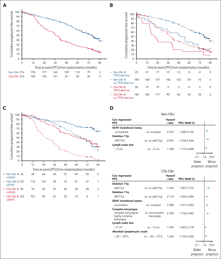
Figure 1.
PFS. (A) PFS according to Ven-Obi (blue) and Clb-Obi (red) arm. (B) PFS according to presence (solid line) or absence (dashed line) of TP53 deletion/mutation. (C) PFS according to mutated- (solid line) or unmutated- (dashed line) IGHV status. (D) Multivariable models for PFS for the Ven-Obi (upper panel) and Clb-Obi arm (lower panel). del/mut, deletion and/or mutation; mIGHV, mutated IGHV; uIGHV, unmutated IGHV.

