FIGURE 4.

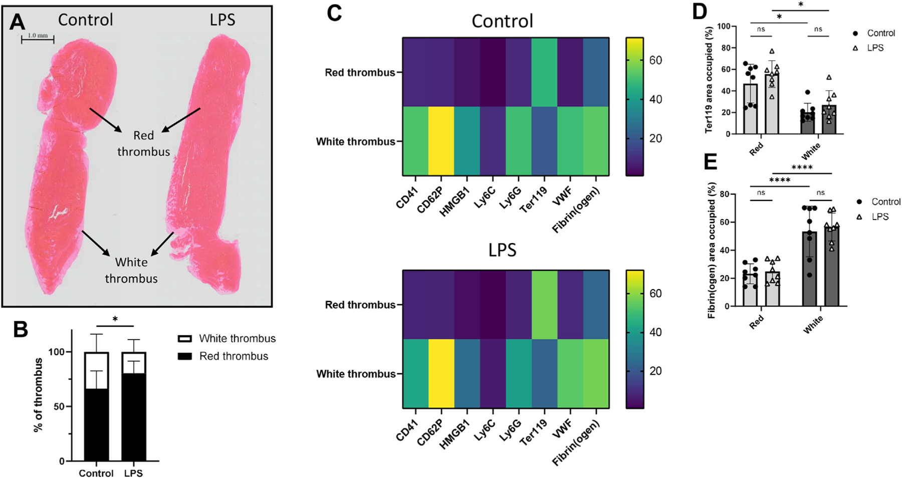
Endotoxemia significantly increases the proportion of the red thrombus, but most thrombus components largely localize in the white thrombus. (A) Hematoxylin and eosin stains of thrombi obtained from control (left) and LPS- (right) treated mice at 24 hours post-stenosis show distinct red and white thrombi. Images are representative of thrombi from n = 7 control and n = 12 LPS mice. Scale bar, 1.0 mm. (B) Mean areas of red and white thrombi as percentages of the entire thrombus in control and LPS cohorts. (C) Heatmaps representing the areas occupied by CD41 (platelets), CD62P (P-selectin), HMGB1, Ly6C (monocytes), Ly6G (neutrophils), Ter119 (RBCs), VWF, and fibrin(ogen) as percentages of the red and white thrombi in the control (top) and LPS (bottom) cohorts. The scale on the right represents percentages. Areas occupied by (D) Ter119 and (E) fibrin(ogen) in thrombi derived from control and LPS-treated mice as percentages of red and white thrombi. *p < .05, ****p < .0001. LPS, lipopolysaccharide; ns, not significant; VWF, von Willebrand factor; VWF:Ag, plasma von Willebrand factor; VWF:CB, von Willebrand factor–collagen binding activity.
