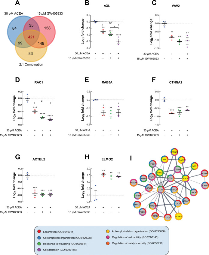
Fig 7. The cell locomotion-related proteomic signature profiles of MDA-MB-231 after exposed CB agonists were investigated.
(A) Venn diagram comparing among 30 μM ACEA, 15 μM GW405833 and 2:1 combination. The dot plots represented protein expression as log2 fold change of CB agonists treated group normalized to control in MDA-MB-231;(B) AXL; (C) VAV2; (D) RAC1; (E) RAB5A; (F) CTNNA2; (G) ACTBL2; (H) ELMO2; (I) Protein-protein interaction diagram of cell locomotion related protein in 2:1 combination treatment. All dot plots were presented as individual log2 fold change ± SEM of three biological replicates. (*p < 0.05, **p < 0.01, ***p < 0.001, ****p < 0.0001 versus control, while #p < 0.05, ##p < 0.01).

