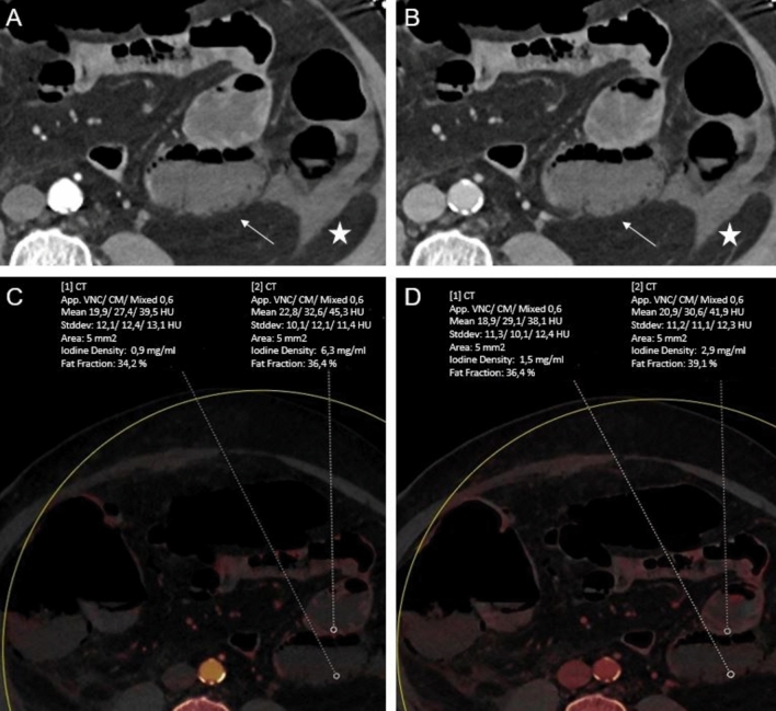
Fig. 4.
60-year-old woman with acute abdomen and elevated lactate. Conventional transversal late arterial (LA, A) and portal venous (PV, B) demonstrate free intraabdominal fluid (star) next to a small bowel loop (arrow). Corresponding dual-energy CT iodine maps based on LA and PV phase (C and D, respectively) demonstrate a greater difference in iodine concentration (IC) reduction of this small bowel loop compared with adjacent segments in the LA phase (LA: 0.9 vs 6.3 mg/ml; PV: 1.5 vs 2.9 mg/ml), indicating significant ischemia which was proven by surgery. CT value measurements showed only a slight reduction (LA: 39.5 vs 45.3; PV: 38.1 vs 41.9)

