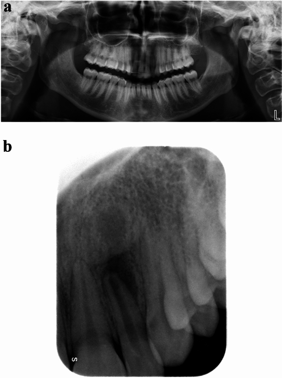
Fig. 4.

Apical periodontitis affecting the upper left lateral incisor. A In panoramic radiography, no periapical bone lesion was detected at the level of the periapex. A large area of radiolucency around incisors of both sides and especially around the root of both lateral incisors can be observed because of the overlap of the air inside the nasal cavity. B Same patient. In periapical radiography, changes in bone structure with clear mineral loss can be undoubtedly noticeable at the level of the periapex
