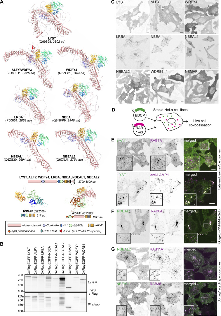
Figure 1.
Mapping the cellular localization of BDCPs, a family of large cytosolic alpha-solenoid/beta-propeller domain proteins. (A) Alpha-fold models and schematic structures of typical, atypical, and small BDCPs. (B) Western blot and anti-flag immunoprecipitation of cell lysates from HeLa cells stably expressing 3xFlag-EGFP-tagged full-length human BDCPs. (C) Representative confocal images of HeLa cells stably expressing 3xFlag-EGFP-tagged human BDCPs. Scale bars 10 µm. (D) Schematic of the live-cell imaging screen done to characterize the nature of BDCP-positive compartments. HeLa cells stably expressing a single BDCP and one of 42 RAB small GTPase fused to 3xFlag-EGFP and mScarlet, respectively, were imaged live using a spinning-disc confocal microscope to detect colocalization and co-migration of both proteins. See Table 1 for an overview of the screen results. (E) HeLa cells stably expressing tdNG-LYST and mScarlet-RAB7A were imaged live (upper panel) or fixed and stained with an anti-LAMP1 antibody (lower panel). Scale bars 10 µm (main figure) or 1 µm (inset magnifications). (F) HeLa cells stably expressing 3xFlag-EGFP-NBEAL1 and mScarlet-RAB6A were imaged live. Scale bars 10 µm (main figure) or 1 µm (inset magnifications). (G) HeLa cells stably expressing 3xFlag-EGFP-NBEAL2 and mScarlet-RAB11A (upper panel) or -RAB38 (lower panel) were imaged live. Scale bars 10 µm (main figure) or 1 µm (inset magnifications). Source data are available for this figure: SourceData F1.

