Figure 6.
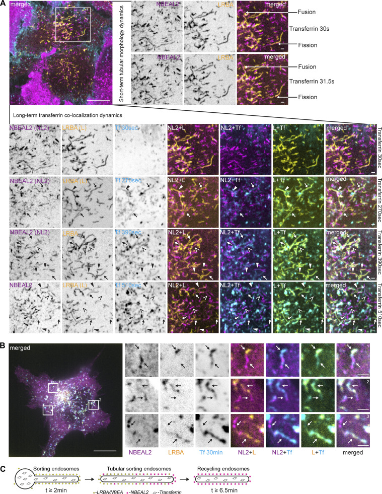
NBEAL2 shows slower dynamics of colocalization with endocytosed transferrin. (A) HeLa cells stably transfected with 3xFlag-EGFP-LRBA and mScarlet-NBEAL2 were imaged live with 1-min intervals starting 30 s after supplementation of growth media with 10 µg/ml of AlexaFluor647-Transferrin. Tubules positive for LRBA and NBEAL2 undergo fast fusion or fission events (right zoomed-in inserts). LRBA-positive tubules that accumulate transferrin between 2.5 and 6 min after its addition to the media are either negative (arrow) or only weakly positive for NBEAL2 (solid arrowhead). Tubular structures positive for NBEAL2 and transferrin, but negative for LRBA (open arrowheads), could be observed starting from 6.5 min after the addition of transferrin to the culture media (lower zoomed-in inserts) Scale bars 10 µm (main figure) or 1 µm (inset magnifications). (B) HeLa cells stably transfected with 3xFlag-EGFP-NBEAL2 and mScarlet-LRBA were treated for 30 min with 10 µg/ml of AlexaFluor647-transferrin in complete media, fixed, and imaged by confocal microscopy. LRBA and NBEAL2 localize to opposite ends of the same transferrin-positive tubules. Scale bars 10 µm (main figure) or 1 µm (inset magnifications). (C) Schematic summary of data from Fig. 6, A and B.

