Figure S1.
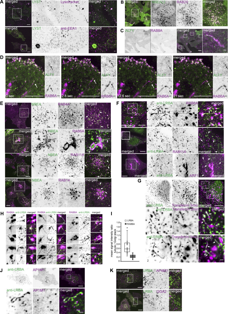
Mapping the cellular localization of BDCPs. (A) HeLa cells stably transfected with LYST tagged with a tandem dimer NeonGreen were treated for 30 min with 20 nM of Lysotracker Deep Red and imaged live (upper panel) or fixed and stained with an anti-EEA1 antibody (lower panel). Scale bar 10 µm (left merged) or 1 µm (right merged). (B) HeLa cells stably transfected with 3xFlag-EGFP-NBEAL2 and mScarlet-I-RAB25 were imaged live. Scale bar 10 µm (left merged) or 1 µm (right merged). (C) HeLa cells stably transfected with 3xFlag-EGFP-ALFY and mScarlet-I-RAB8A were imaged live. Scale bar 10 µm (left merged) or 1 µm (right merged). (D) HeLa cells stably transfected with 3xFlag-EGFP-ALFY and mScarlet-RAB6A were imaged live. ALFY speckle co-migrates with the tip of RAB6A-positive tubule (arrows). Scale bar 10 µm (main figure) or 1 µm (inset magnifications). (E) HeLa cells stably transfected with 3xFlag-EGFP-NBEA and mScarlet tagged RAB4A, RAB6A, RAB11A, or RAB14 were imaged live. NBEA partially colocalizes with the tubular segments of RAB4A-, RAB11A,- or RAB14- positive endosomes and RAB6A-positive Golgi-connected tubules (arrows). Scale bar 10 µm (left merged) or 1 µm (right merged). (F) HeLa cells stably transfected with the indicated small GTPases were fixed and stained with an anti-LRBA antibody. Endogenous LRBA colocalizes with mScarlet-RAB6A, mScarlet-RAB11A and ARF1-mScarlet on cytosolic tubules (arrows). Scale bar 10 µm (left merged) or 1 µm (right merged). (G) HeLa cells fixed 3 min after the addition of 10 µg/ml of AlexaFluor647-Transferrin to the culture media and stained with an anti-LRBA antibody. Scale bar 10 µm (upper merged) or 1 µm (middle and lower merged. (H) Examples of mScarlet-RAB6A-positive Golgi and Golgi-connected tubules that colocalize with endogenous LRBA as in F upper panel. Scale bars 1 µm. (I) Quantification of the mean signal intensity ratio of LRBA or RAB6A signals on LRBA/RAB6A positive tubule vs. nearby LRBA/RAB6A positive Golgi stack. The mean signal intensity of green (endogenous LRBA) or magenta (mScarlet-RAB6A) channels within a 4 × 4 pixels rectangular box over the tubule or Golgi stack was used for calculations. n = 20. (J) HeLa cells stably transfected with AP1M1-mScarlet stained with an antibody against endogenous LRBA. Scale bar 10 µm (upper merged) or 1 µm (lower merged). (K) HeLa cells stably transfected with 3xFlag-EGFP-LRBA and AP4M1-mScarlet or GGA2-mScarlet imaged live. Scale bars 10 µm (left merged) or 1 µm (right merged).

