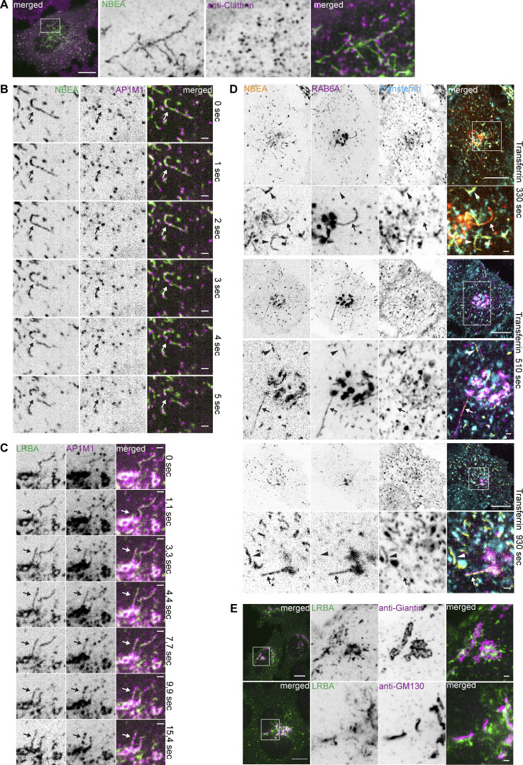
Figure S2.
Characterization of LRBA- and NBEA- positive compartments. (A) HeLa cells stably transfected with 3xFlag-EGFP-NBEA were fixed and stained with an anti-clathrin antibody. Scale bar 10 µm (left merged) or 1 µm (right merged). (B) HeLa cells stably transfected with 3xFlag-EGFP-NBEA and AP1M1-mScarlet were imaged live with 1 s intervals. NBEA- and AP1M1-positive tubules undergo rapid fission at or close to AP1M1-positive speckles (arrow). Scale bars 1 µm. (C) Live cell imaging of HeLa cells stably transfected with 3xFlag-EGFP-LRBA and AP1M1-mScarlet. LRBA and AP1M1 into distinct subdomains along the length of the growing tubule (arrows). Scale bars 1 µm. (D) HeLa cells stably transfected with 3xFlag-EGFP-NBEA and mScarlet-RAB6A were imaged live with 1 min intervals starting 30 s after supplementation of growth media with 10 µg/ml of AlexaFluor647-Transferrin. NBEA-decorated tubules, positive for RAB6A (arrow) do not accumulate transferrin (arrowhead). Scale bars 10 µm (main figures) or 1 µm (zoomed in inserts). (E) HeLa cells stably transfected with 3xFlag-EGFP-LRBA were fixed and stained with anti-Giantin (upper panel) or anti-GF130 (lower panel) antibodies. Endogenous markers of media-Golgi (giantin) and cis-Golgi (GM130) localize in close proximity to perinuclear 3xFlag-EGFP-LRBA positive structures but do not colocalize with LRBA-positive tubules. Scale bars 10 µm (left merged) or 1 µm (right merged).

