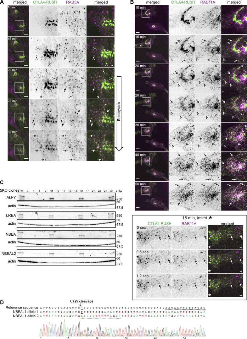
Figure S4.
Characterization of intracellular trafficking of CTLA4-EGFP-RUSH protein and confirmation of BDCPs knockouts. (A) Live cell imaging of HeLa cells stably transfected with mScarlet-RAB5A and transiently transfected with CTLA4-EGFP-RUSH at the indicated time points after the addition of 50 µM biotin. Arrows and open arrowheads point to colocalization or lack of colocalization of CTLA4 with RAB5A, respectively. Scale bars 10 µm (left merged) or 1 µm (right merged). (B) Live cell imaging of HeLa cells stably transfected with mScarlet-RAB11A and transiently transfected with CTLA4-EGFP-RUSH at the indicated time points after the addition of 50 µM biotin. Arrows and open arrowheads point to colocalization or lack of colocalization of CTLA4 with RAB11A, respectively. The box labeled with an asterisk (16 min, insert*) is zoomed in in the lower subpanel to demonstrate colocalization of a fraction of CTLA4 with RAB11A at the 16 min time point. Scale bars 10 µm (left merged) or 1 µm (right merged). (C) Western blot of clones of HeLa cells with knockout (5KO) of the reference isoforms of ALFY, LRBA, NBEA, NBEAL1, and NBEAL2. Clones 8 and 12 were renamed as 5KO1 and 5KO2 and used for surface biotinylation experiments. (D) Genotyping of HeLa cells with knockout of the reference isoform of NBEAL1. Solid line marks the insertion of a single nucleotide in allele 1, while the dotted line highlights remaining homologous sequence after the deletion of 28 nucleotides in NBEAL1 allele 2. Source data are available for this figure: SourceData FS4.

