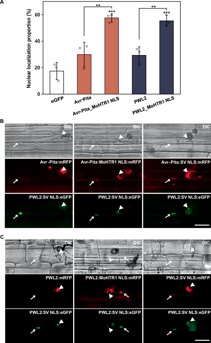
Fig. 3. MoHTR1 NLS leading the nuclear transport of cytoplasmic effectors of M. oryzae in rice protoplasts and rice sheath cells.
A Intracellular localization of Avr-Pita and PWL2, two cytoplasmic effectors of M. oryzae, in rice protoplasts. Each cytoplasmic effector was fused with MoHTR1 NLS, and cloned into eGFP-expressing plasmid under CaMV 35S promoter. The proportion of these cytoplasmic effectors in nuclei was measured under a fluorescence microscope. B Localization of Avr-Pita and Avr-Pita tagged with a positive control NLS (SV NLS) and MoHTR1 NLS in rice sheath cells. Avr-Pita with mRFP and NLS were expressed under Avr-Pita native promoter. PWL2:eGFP:SV NLS was used as biotrophic interfacial complex (BIC) and rice nuclei marker. Arrowheads indicate BICs and arrows indicate rice nuclei. Scale bar; 20 μm. C Localization of PWL2 and PWL2 tagged with SV NLS and MoHTR1 NLS in rice sheath cells. PWL2 and PWL2 with SV NLS and MoHTR1 NLS were expressed under PWL2 native promoter and transformed into the wild type of M. oryzae. PWL2:eGFP:SV NLS was used as BIC and rice nuclei marker. The localization of Avr-Pita and PWL2 was observed at 30–34 hpi. Arrowheads indicate BICs and arrows indicate rice nuclei. Scale bar; 20 μm. Mean ± SD, n = 3 independently transfected protoplasts, significance was determined by an unpaired two-tailed Student’s t-test (**p < 0.01 and ***p < 0.001). Representative data are shown from independently experiments and source data are provided as a Source Data file.

