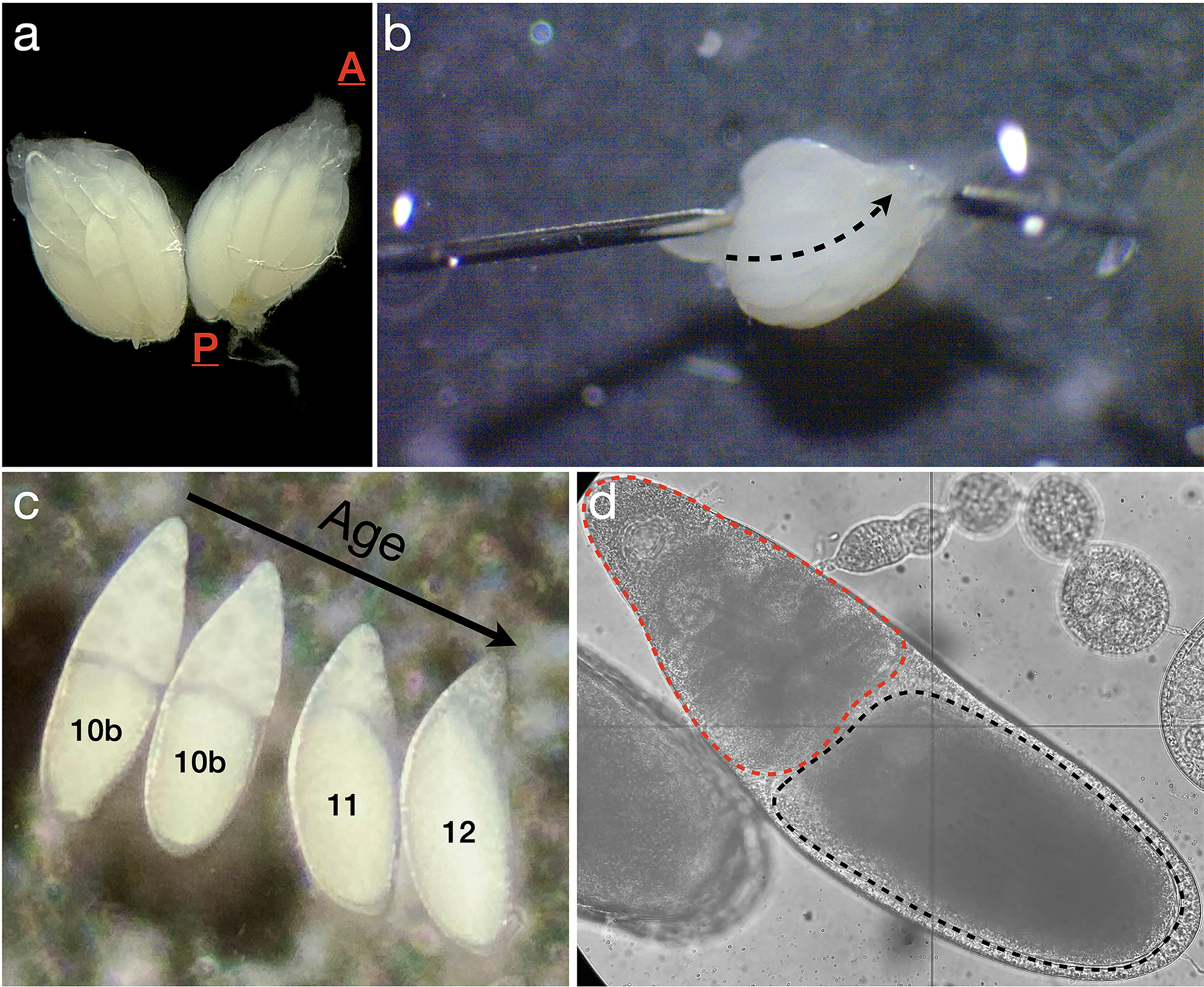
Figure 2: Ovary dissection and identification of egg chambers undergoing nurse cell dumping.

A. One pair of ovaries removed from a fruit fly; A is the anterior end, where the germaria are located, and P is the posterior end, where mature stage 14 eggs reside. B. Disruption of the peritoneal muscle sheath. The needle on the left is used to immobilize the ovary, while the needle on the right is moved gently along the dashed arrow, between ovarioles, to remove the muscle sheath holding the ovarioles together. C. Four egg chambers in stages 10b-12, with the youngest (stage 10b) just beginning the nurse cell dumping process at left, and the oldest (stage 12) shortly after dumping is completed at right. D. Brightfield image of an egg chamber approximately 10–20 minutes into the dumping process with the oocyte (black dashed outline) and the nurse cell cluster (red dashed outline) highlighted.
