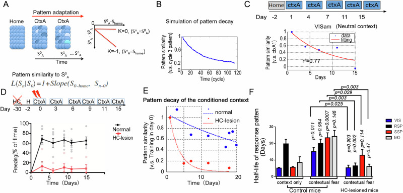
Fig. 4. Hippocampus reinstates cortical representation pattern in multiple areas of neocortex for the conditioned context.
A, B Simulation of the pattern similarity decay using the empirical parameters of the population plasticity. A Gaussian-distributed activity pattern was used as the initial seed. The pattern in the third cycle was treated as the initial representation. α, is the slope parameter. C Representative example of pattern similarity decay after repeated CtxA exposure in VIS cortex of mice. The mouse explored the box A for 3 min each day. Data was fitted with an exponential curve (red). D Freezing in box A after contextual fear conditioning in hippocampus-lesion group (n = 5 mice) and normal group (n = 9 mice), Two-way ANOVA test, group p < 0.0001. E Representative example of population activity pattern decay in the VISam of normal mice (Blue) and HC-lesion mice (Red). F Quantifications of the half-life for the pattern decay in each recorded areas of normal mice (Control) and HC-lesion mice. Context only group, n = 6 mice; contextual fear group, normal, n = 9 mice; HC-lesion, n = 5 mice. Multiple cortices were recorded from one mouse. The roof of half-life was set to 40 days. Error bars, s.e.m.

