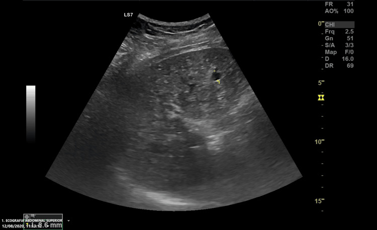
Figure 1. Abdominal Ultrasound Image.
Abdominal ultrasound revealing a liver with diffusely heterogeneous parenchyma and multiple indeterminate hypoechoic foci (the biggest one in this image measured and labeled with the number 1). This initial imaging study raised the suspicion of Von-Meyenburg complexes.

