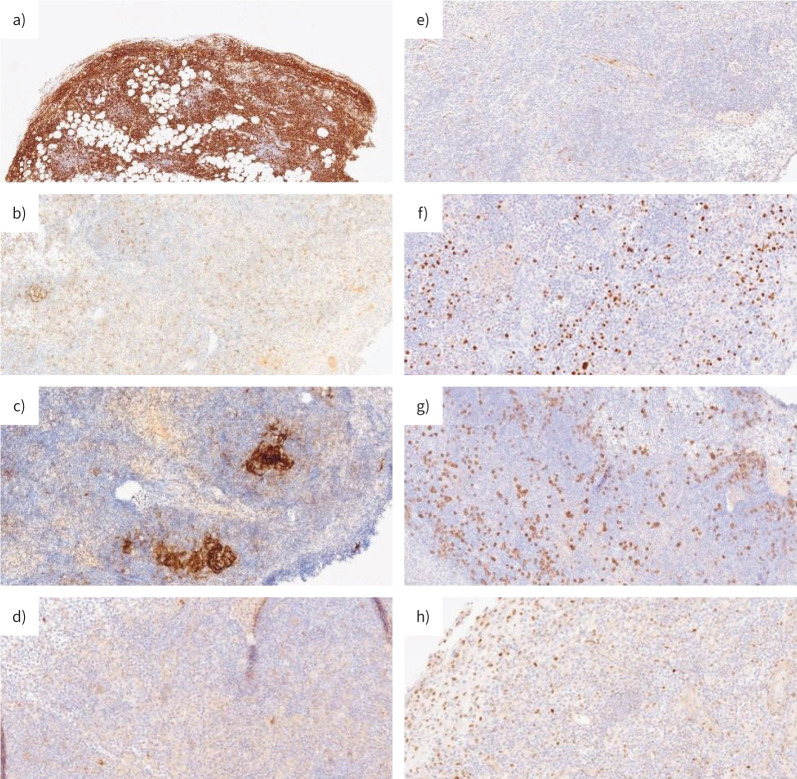
FIGURE 5.
Immunohistochemical analysis of the lymphoid component of the lesion. a) Lymphoid cells were CD20 positive. CD20 stain demonstrates B-cell origin of the lymphoid diffuse infiltrate in pleural and subpleural tissue. Lymphoid cells were negative for b) CD21; c) CD10; d) CD23; e) Cyclin D; f) MUM-1; g) CD138; h) BCL-6 protein expression.

