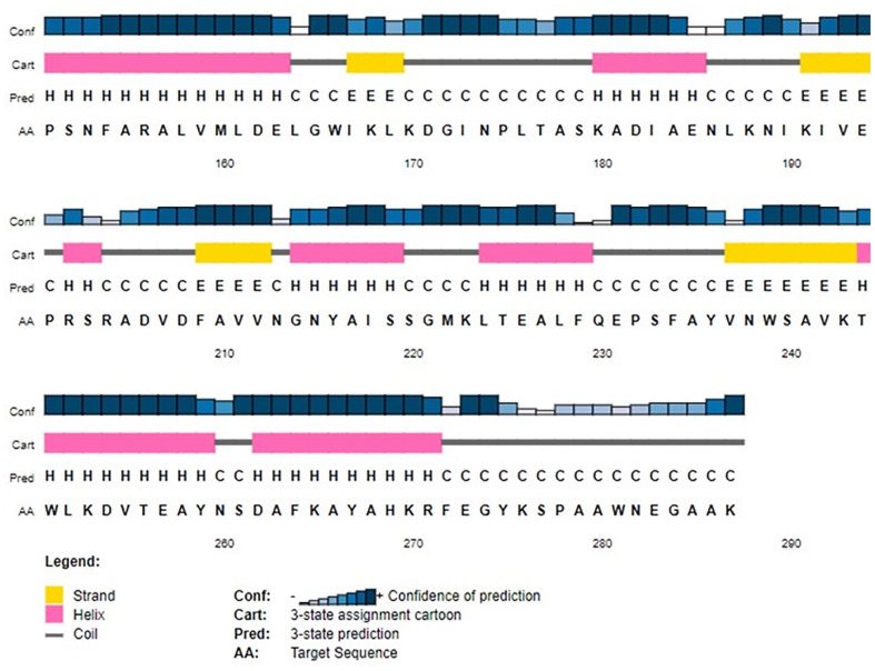
Figure 5.
Secondary structure of HP PBJ89160.1.
PSI-PRED server predicted the target protein’s secondary structure. Four distinct components make up this graphic illustration. Bars in the first part are varied heights. The confidence score is proportional to the length of the bar height. In the second section, the alpha helix is represented by the pink color, the beta sheets or strands are represented by the yellow color, and the coils are represented by the gray color. A coil links a specific beta sheet to a specific alpha helix. The secondary structure of a protein is depicted alphabetically in the third section; here, the letters E, H, and C stand in for beta sheets, alpha helixes, and coils, respectively. The order of amino acids is listed alphabetically in the final section.

