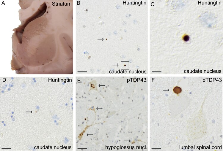
Figure 2.
Histopathological findings of huntingtin and pTDP-43 inclusions in MND patient #1, carrying reduced penetrance HTT repeat expansion (HTT 36/19). A coronal section of a fresh brain showing the striatum (A) that has regular size and no atrophy. Staining with an antibody against huntingtin shows small extranuclear huntingtin staining (arrows) in the caudate nucleus (B), enlarged in (C). The area of huntingtin staining was small, and huntingtin was located in the extranuclear space (D). Using the pTDP-43 antibody against phosphorylated TDP-43, cytoplasmic TDP43 inclusions are seen in the motor neurons of nucleus hypoglossus (E) and large granular cytoplasmic pTDP43-inclusions were observed in lumbar spinal cord (F). Scale bar represents 100 μm in b-f.

