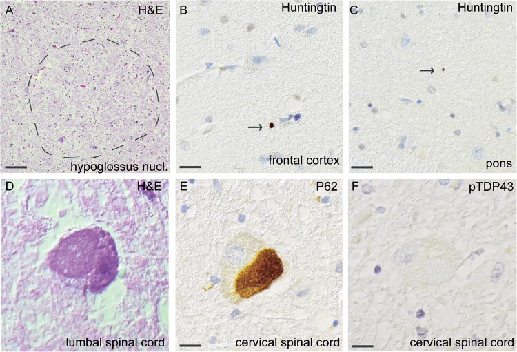
Figure 4.
Histopathological findings of huntingtin and p62 inclusions, but no pTDP43-inclusions, in MND patient #3, carrying HTT repeat expansion within intermediate range (HTT 33/22). Loss of neurons in the hypoglossus nucleus stained with H&E (A). Extranuclear small inclusions of huntingtin were seen in the frontal cortex (B) and in pons (C). Swollen eosinophilic motor neuron in lumbar spinal cord stained with H&E (d). Note the swollen eosinophilic appearance. Staining with an antibody against p62, large round p62-positive inclusions are observed in the cervical spinal cord (E). No inclusions are seen in the cervical spinal cord in when stained with pTDP-43 (f). Scale bar represents 20 μm in a, 100 μm in b-f.

