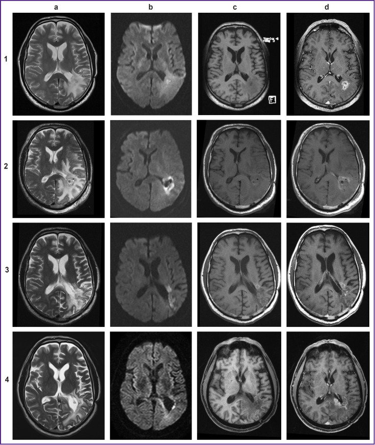
Figure 4. MRI of the brain with contrast intensifying:
1 — before intervention: (a) T2-weighted image (WI) before intervention; (b) diffusion-weighted images (DWI) (b=1000); (c) T1-WI before contrast; (d) post-contrast T1-WIs;
2 — after stereotactic photodynamic therapy: (a) T2-WI, increase in the perifocal area of edema; (b) DWI (b=1000), limitated diffusion in the affected area; (c) T1-WI before contrast, a minor increase in signal intensity in the affected area; (d) T1-WIs post-contrast, decreased accumulation of the contrast agent in the affected area;
3 — 6 months after stereotactic photodynamic therapy: (a) T2-WI, reduction in the area of perifocal edema; (b) DWI (b=1000), limitated diffusion in the affected area and anterior to it; (c) T1-WI before contrast, a minor hyperintense area anterior to the treatment area; (d) post-contrast T1-WIs, a minor area of insignificant accumulation of the contrast agent in the affected area;
4 — 14 months after stereotactic photodynamic therapy (continued growth): (a) T2-WI; (b) DWI (b=1000), remaining minor area of a limitated diffusion in the impact area; (c) T1-WI before contrast; (d) post-contrast T1-WIs, the affected area still has a insignificant accumulation of the contrast agent and a new area of accumulation of the contrast agent measuring 38×36×33 mm

