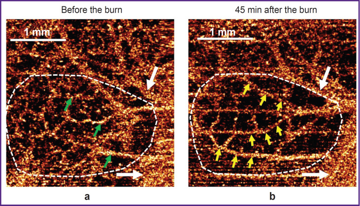
Figure 3. Example of disappearance of small-caliber blood vessels and simultaneous activation of shunt vessels identified in OCTA images of the colon wall 45 min after thermal burn induction (the region of comparison is designated by a white dashed line):
(a) before the burn; (b) after the burn induction; white arrows — large vessels identical in both images are visualized similarly before and after the burn; green arrows — inactive shunt vessels; yellow arrows — active shunt vessels

