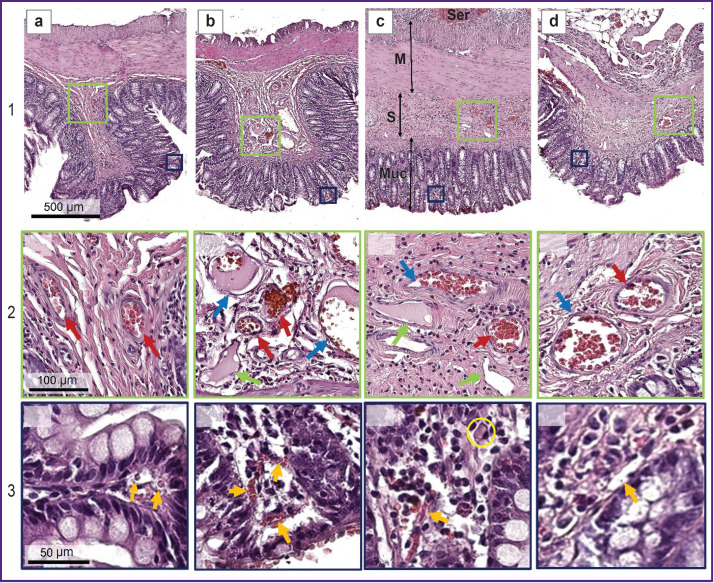
Figure 5. Histological images of the colon wall before burn induction (a), 45 min after (b), on day 7 (c), and on day 14 (d) after the induction of thermal injury:
1 — overview images, the colon wall consists of four layers: a thin serosa (Ser), double-layered muscularis externa (M), submucosa (S), and mucosa (Muc); the colon folds are formed by mucosa and submucosa (a1), (b1);
2 — magnified images of the submucosa with vessels; red arrows — arterioles, blue arrows — venules, green arrows — lymphatic vessels;
3 — magnified images of the mucosa and lamina propria; orange arrows — capillaries, a hyaline thrombus in the capillary lumen is designated with a yellow circle

