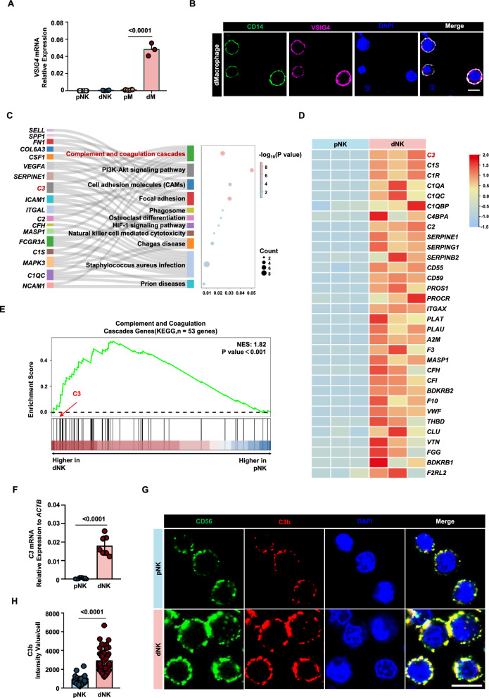
Fig. 4.
dNK cells from normal pregnancy express the natural ligand of VSIG4. A Gene expression levels of VSIG4 in peripheral blood NK cells (pNK), peripheral blood monocytes (pM), decidual macrophages (dM), and decidual NK cells (dNK). B Fluorescence image depicting the expression of VSIG4 protein on decidual macrophages. C Biological processes that are significantly enriched in Sankey dot pathway enrichment analysis of the upregulated genes of dNK relative to pNK. D Heat map of differential genes in complement and coagulation cascade pathways, n = 3 per group. E Gene set enrichment analysis (GSEA) revealed an increase in complement and coagulation cascade pathways (enrichment plot: COMPLEMENT AND COAGULATION CASCADES PATHWAYS, HSA04610) in dNK cells compared with pNK cells, n = 3 per group. F qPCR validation of the differential expression of the C3 gene in dNK cells and pNK cells. G CLSM images showing the secretion of C3b in pNKs and dNKs. The images were captured using the same parameters. Scale bars = 5 μm. H Statistical histogram of C3b mean fluorescence intensity of each decidual or peripheral NK cell. P values have been determined by two-tailed unpaired t-test

