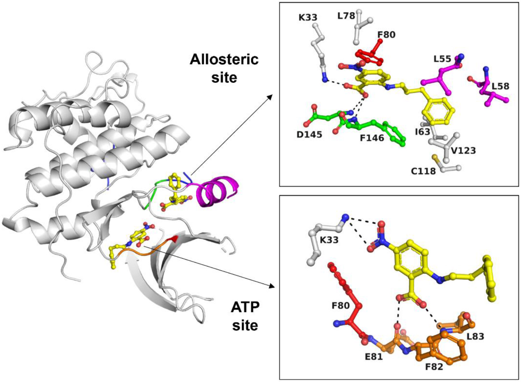
Fig. 2. Hit compound NPPB binds to an allosteric pocket in CDK2.

Hit NPPB binds to both the ATP site and the adjacent allosteric pocket of CDK2 (PDB ID 7RWE). For the allosteric site, all residues in the immediate vicinity of NPPB (d < 4 Å) are shown. For the ATP site, only the hinge region and Lys33 are shown. The different colors denote the DFG motif (green), the C-helix (magenta), the gatekeeper residue (red), the hinge region (orange) and the activation loop (blue). H-bonding and polar interactions are indicated as black dotted lines.
