Fig. 7.

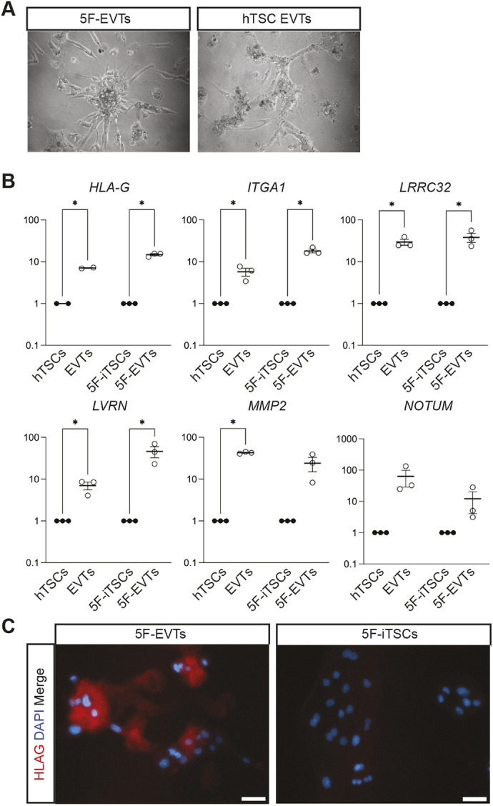
5F-iTSCs can be directed to differentiate to extravillous trophoblast. (A) Brightfield imaging of EVTs differentiated from 5F-iTSCs and hTSCs. (B) RT-qPCR analysis for the detection of the selected EVT markers HLA-G, ITGA1, LRRC32, LVRN, MMP2 and NOTUM. Relative expression is shown as fold change over undifferentiated 5F-iTSCs normalised to GAPDH. Data are mean±s.e.m. of n=3 biological replicates analysed with an unpaired one-tailed t-test (*P<0.05). (C) Immunofluorescence analysis for the detection of HLA-G (red) and DAPI nuclear staining (blue) in 5F-EVTs and 5F-iTSCs. Scale bars: 50 μm.
