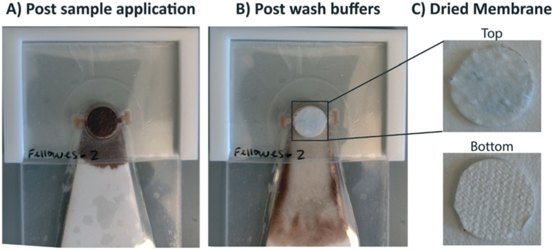
Fig. 3.

Demonstration of SNAPflex with whole blood samples. A) Application of lysed, precipitated blood (100 μL blood sample input). B) Purified capture membrane after application of three wash buffers. C) The top and bottom of the capture membrane shows significant removal of blood components, with blue glycogen precipitant remaining on top of the capture membrane.
