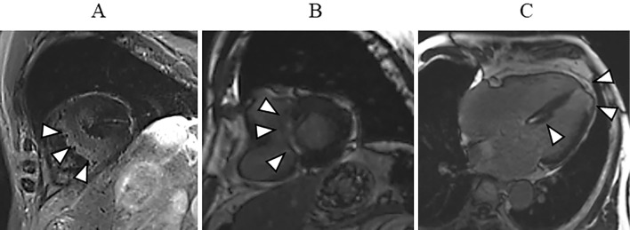
Figure 4.
In cardiac magnetic resonance imaging, a T2-weighted black-blood image showed high intensity in the interventricular septum (arrowheads) (A). Delayed gadolinium enhancement was seen in the middle layer of the interventricular septum (B, arrowheads; C, arrowheads), and subendocardial wall of the left ventricular apex (arrowheads) (C).

