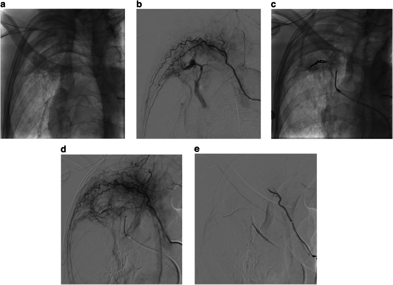
Fig. 5.
a Right anterior oblique control image of the right upper chest showing right apical scarring and a mycetoma. b DSA of a right upper intercostal artery demonstrating marked systemic to pulmonary artery shunting and a pulmonary artery branch pseudoaneurysm. c and d Control image, followed by an intercostal arteriogram following coil embolization from the pulmonary arterial side demonstrating occlusion of the pulmonary artery. e Completion DSA following embolization of the intercostal artery with 150–250 µm PVA particles obliterating systemic to pulmonary artery shunting

