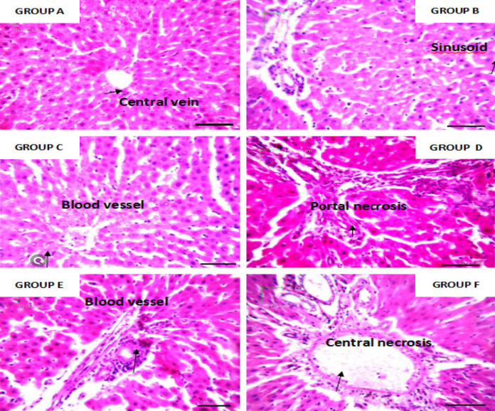
Figure 9.
Photomicrograph of liver tissue of adult rats stained with hematoxylin and eosin. High concentration of both leaf extracts shows morphological changes with inflammatory cell infiltrates while the others show normal histology. ×400 magnification and scale bar = 50 um. Conclusion: Both extracts show hepatotoxicity at high concentrations.

