FIGURE 1.

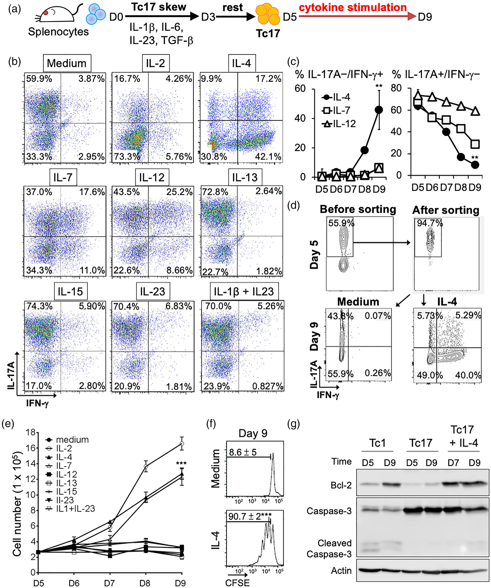
Effects of different cytokines on conversion and expansion of Tc17 cells. (a) Schema of Tc17 cells polarization and cytokine stimulation. Splenocytes were incubated in Tc17 skewing medium for 3 days (D3). After 2 days’ rest, different cytokines were added to Tc17 cells for further 4 days (D9). (b) Representative dot plots showing population changes in IL17A- and IFN-γ-producing cells following stimulation with different cytokines at day 9 (D9). (c) Average changes in IL-17A/IFN-γ population upon IL-4, IL-7, or IL-12 stimulation. Mean ± SD are shown form three independent experiments. **P < 0.01. (d) IL-4 treatment of IL-17A producing cells programs them into IFN-γ-producing cells. Cytokine secretion assay was used to label and sort IL-17A producing cells. Cells were treated with or without IL-4 for 4 days. Representative results were shown from one out of three independent experiments. (e) Effects of different cytokines on Tc17 expansion. Viable cells were counted using trypan blue exclusion assay. Mean ± SD of three independent experiments was shown. (f) IL-4 enhanced Tc17 proliferation. CFSE-labelled assay was used to measure proliferation of Tc17 cells. Mean ± SD of three independent experiments was shown at right. ***P < 0.001. (g) IL-4 induces anti-apoptosis signature in Tc17 cells. Expression of Bcl-2 and Caspase3 in Tc1 and Tc17 cells was analysed by Western blot. Actin was used as a loading control
