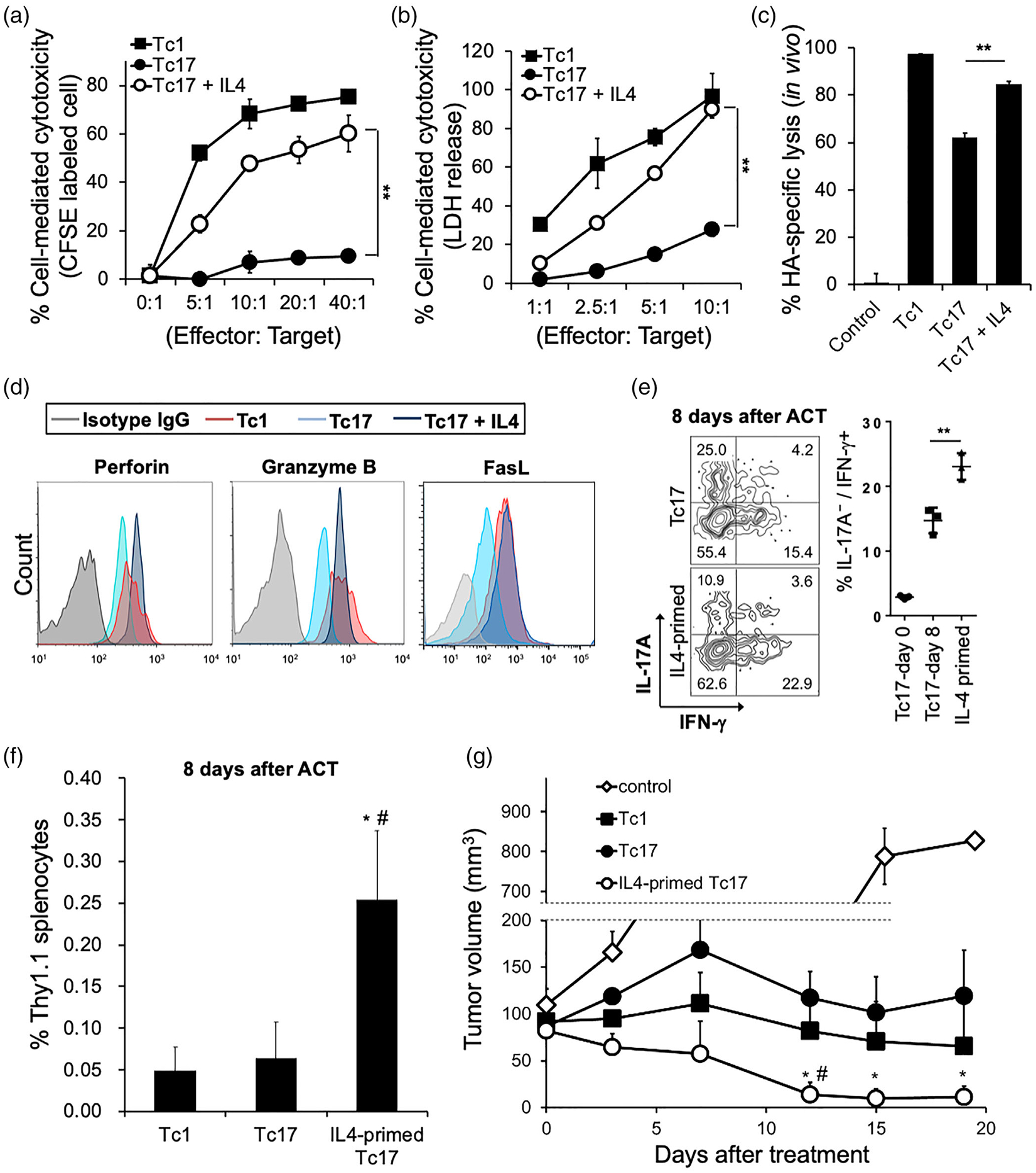
FIGURE 2.

IL-4 enhances the anti-tumour cytotoxicity of Tc17 cells. (a and b) In vitro cytotoxicity of IL-4 programmed Tc17 cells. The HA-specific effectors were polarized under Tc1 and Tc17 skewing conditions. Target cells (CT26-HA) and non-target cells (CT26) were labelled and mixed in different effector: Target ratio for 16 h. Specific lysis of target cells was calculated as (1-target/nontarget) × 100%. Mean ± SD was shown from three independent experiments. **P < 0.01. (b) Target cells (CT26-HA) and control cells were mixed under different effector: Target ratio for 16 h and measured as a 492 nm absorbance. Percentage of specific lysis are average from three independent experiments; mean ± SD was shown. **P < 0.01. (c) In vivo cytotoxicity of IL-4 programmed Tc17 cells. Polarized HA-specific Tc1, Tc17, and IL-4-stimulated Tc17 were injected intravenously into recipient mice. HA-primed target cells and non-target cells were labelled by CFSE, and injected to the recipient mice 6 h later. Specific lysis of target cells was measured after 16 h, and calculated as (1-target/nontarget) × 100% (n = 4). Mean ± SD was shown. **P < 0.01. (d) Expression of Perforin, Granzyme B, and Fas-ligand (FasL) in polarized HA-specific Tc1, Tc17, and IL-4-stimulated Tc17 cells. One representative experiment of three with similar results is shown. (e) In vivo conversion of Tc17 cells into IFN-γ-producing cells. HA-specific Tc17 cells (Thy1.1) were stimulated with or without IL-4 for 24 h, then injected into the CT26HA tumour-bearing mice (Thy1.2) for 8 days (n = 3). IL-17A/IFN-γ producing populations of Thy1.1 positive lymphocytes were measured by flow cytometry. Experiments were independently repeated three times with similar results. Mean ± SD is shown. **P < 0.01. (f) Tc cell expansion after adoptive cell therapy. CT26-HA tumour-bearing mice received HA-specific Tc1, Tc17, or IL-4-primed Tc17 cells (1 106), and cell expansion analysed 8 days after cell transfer mean ± SD was shown. *P < 0.05 to Tc17. # P < 0.05 to Tc1. (g) Comparison of the antitumour efficacy of Tc1, Tc17, and IL-4-primed Tc17 in a CT26-HA tumour-bearing model. HA-specific Tc1, Tc17, and IL-4-stimulated Tc17 cells (1× 10⁶) were injected intravenously into tumour-bearing mice (n = 7 per group). Tumour size was monitored serially after ACT. Mean ± SEM was shown. *P < 0.05 to Tc17. #P < 0.05 to Tc1
