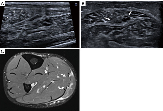
Figure 2.
A 40-year-old woman, with a gastrocnemius aponeurotic injury (type 2A) at 48 hours, after suffering an acute pain of the calf during a tennis match. Long and axial axis US images (A,B) show the perimysium and muscle fascicles are retracted (arrowheads) secondary to an aponeurotic rupture affecting less than 50% of total muscle width (arrows) and a mild hematic intermuscular suffusion. Axial fat-suppressed TSE intermediate-weighted image (C) of the same type of injury shows an aponeurotic rupture affecting 50% of total muscle width (arrows) and feathery edema of the muscle fibers. There is no significant hematomas. See Video 2 showing synchronous motion of the gastrocnemius and soleus muscles. US, ultrasound; TSE, turbo spin-echo.

